


Lung Cancer

Overview
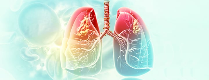
Lung cancer is the growth of abnormal cells in one or both lungs. These cells can multiply rapidly and turn into tumors that interfere with the function of the lungs and, eventually, spread to other parts of the body.
Lung cancer is the second most common kind of cancer diagnosed in the United States, and accounts for nearly a third of all cancer deaths. Most people who get lung cancer were cigarette smokers, but non-smokers get it too. Exposure to radon, asbestos, and secondhand smoke are also risk factors. In some cases, there is no known cause.
One of the challenging aspects of lung cancer is that it may be years before symptoms emerge. By the time it's diagnosed, about half the patients have cancer that's already spread outside the lungs.
How the lungs work
The lung's job is to remove carbon dioxide from the blood and replace it with oxygen. It acts like a pump with every breath you take. The air you breathe comes in through your nose or mouth, and passes though your trachea, or windpipe, into the lungs through two tubes called main stem bronchi. One of the tubes goes to the right lung and the other one to the left lung.
In the lungs, each of the main stem bronchi divide into smaller tubes, called bronchi, and then into even smaller tubes called bronchioles. The bronchioles end in tiny air sacs called alveoli where the exchange of oxygen and carbon dioxide – the gases you breathe – takes place. There are three sections of lung or lobes on the right side of the chest and two sections on the left side.
Types of lung cancer
Cancers that begin in the lungs are divided into two major types – small cell lung cancer and non-small cell cancer. The two types are distinguished by how the cancer cells look under a microscope. Each type of lung cancer grows and spreads differently and calls for different treatment.
Non-small cell lung cancer is more common and generally grows more slowly. There are four main types of this cancer. They are named for the cells in which the cancer develops: squamous cell carcinoma, adenocarcinoma, bronchoalveolar carcinoma and large cell carcinoma.
Small cell lung cancer, sometimes called oat cell cancer, is less common. This type of lung cancer grows more quickly and is more likely to spread to other organs.
Our approach to lung cancer
UCSF's highly experienced thoracic surgeons and thoracic oncologists provide state-of-the-art care for lung cancer. Working with specialists in radiation oncology, pulmonology, pathology and radiology, we offer each patient a precise diagnosis and a tailored treatment plan.
Our thoracic surgery team helped pioneer minimally invasive lung surgery, first with video-assisted surgery and now with robotic surgery. For patients, this translates to shorter hospital stays, less pain and faster recoveries when compared to traditional procedures. We have established a reputation for accepting complex, challenging cases.
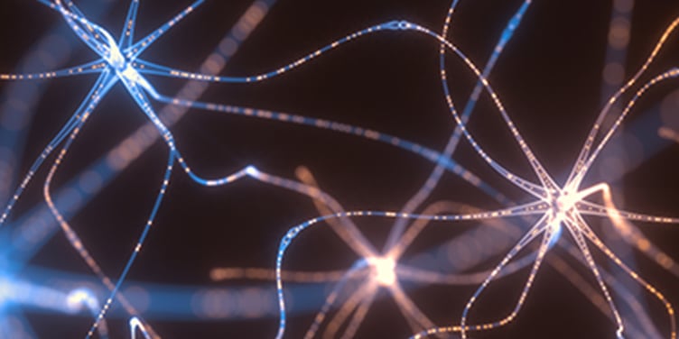
Lung Cancer Mutations and Treatment Advances
KRAS is one of the common genetic mutations that cause lung cancer. Watch leading researchers discuss new treatments on the horizon that target this mutation.
Awards & recognition
-

Among the top hospitals in the nation
-

Best in Northern California and No. 7 in the nation for cancer care
-

No. 8 in the nation for pulmonology & lung surgery
-

Rated high-performing hospital for lung cancer surgery
Signs & symptoms
Common signs and symptoms of lung cancer include:
- A cough that doesn't go away and gets worse over time
- Constant chest pain
- Coughing up blood
- Shortness of breath, wheezing or hoarseness
- Repeated problems with pneumonia or bronchitis
- Swelling of the neck and face
- Loss of appetite or weight loss
- Fatigue
Diagnosis
To help find the cause of symptoms, your doctor will evaluate your medical history, smoking history, exposure to environmental and occupational substances, and family history of cancer. Your doctor will perform a physical exam and may recommend a chest X-ray, computed tomography (CT) scan and other tests.
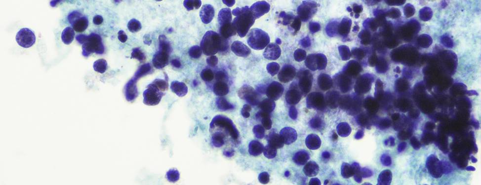
If lung cancer is suspected, sputum cytology – the microscopic examination of cells obtained from a deep-cough sample of mucus in the lungs – is a simple test that may be useful in detecting lung cancer. To confirm the presence of lung cancer, your doctor must examine tissue. A biopsy – the removal of a small sample of tissue for examination under a microscope by a pathologist – can determine if you have cancer. A number of procedures may be used to obtain this tissue:
- Bronchoscopy. A bronchoscope, a thin, lighted tube, is put into your mouth or nose and down your windpipe to look into the breathing passages. Through this tube, your doctor can collect cells or small samples of tissue.
- Needle aspiration. A needle is inserted through the chest into the tumor to remove a sample of tissue.
- Computerized tomography (CT) guided biopsy. A computerized axial tomography scan is more commonly known by its abbreviated name, CAT scan or CT scan. It is an X-ray procedure that combines many X-ray images with the aid of a computer to generate cross-sectional views and, if needed, three-dimensional images of the internal organs and structures of the body. A large, donut-shaped X-ray machine takes images at many different angles in the chest. These images are processed by a computer to create cross-sectional pictures. In each of these pictures the body is seen as an X-ray "slice" of the body, which is recorded on film. This recorded image is called a tomogram. A CT scan is used to define normal and abnormal structures in the body. During a CT-guided biopsy, the scan is used to help the doctor accurately guide the needle into the suspected tumor so a sample can be removed.
- Thoracentesis. This is the removal of fluid in the chest, which can be done under the guidance of ultrasound or CT guidance (see above). Using a needle, the doctor removes a sample of the fluid that surrounds the lungs to check for cancer cells.
Staging
If the diagnosis is cancer, your doctor will determine the stage or extent of the disease. Staging is done to find out whether cancer has spread and, if so, to what parts of the body. Lung cancer often spreads to the brain, bone, liver and adrenal gland. Knowing the stage of the disease will help your doctor plan treatment. Some tests used to determine if cancer has spread include:
- Computerized axial tomography (CAT or CT) scan. A computerized axial tomography scan is more commonly known as a CAT or CT scan. It is a X-ray procedure that combines many X-ray images with the aid of a computer to generate cross-sectional views and, if needed, three-dimensional images of the internal organs and structures of the body. A large, donut-shaped X-ray machine takes images at many different angles in the body. These images are processed by a computer to produce cross-sectional pictures. In each of these pictures the body is seen as an X-ray "slice" of the body, recorded on a film. This recorded image is called a tomogram. A CT scan is used to define normal and abnormal structures in the body.
- Magnetic resonance imaging (MRI). MRI uses radio waves, a powerful electromagnet and a computer to view detailed areas of the body, including the brain.
- Bone scan. A bone scan can show if cancer has spread to the bones. A small amount of radioactive substance is injected into a vein. It travels through the bloodstream and collects in areas of abnormal bone growth. An instrument called a scanner measures the radioactivity levels in these areas and records them on X-ray film. There are no dietary restrictions for this test. The patient usually is given an injection of radioactive tracer that is "tagged" to a calcium-like material about three hours before the scan to give the calcium time to circulate and be taken up by the bone. The patient is then allowed to leave and return to the scanning facility approximately three hours later to complete the scan.
- Positron emission tomography (PET) scan. This imaging technique monitors metabolic, or biochemical, activity in the brain and other organs by tracking the movement and concentration of a radioactive tracer injected into the bloodstream. The technique uses special computerized imaging equipment and rings of detectors surrounding the patient to record gamma radiation produced when positrons (positively-charged particles) emitted by the tracer collide with electrons. It produces images that can be used to measure many vital processes, including glucose metabolism, blood flow and perfusion, and oxygen utilization. With these images, your doctor can identify normal and abnormal states.
- Mediastinoscopy and mediastinotomy. A mediastinoscopy can help show if cancer has spread to the lymph nodes in the chest. Using a lighted viewing instrument, called a scope, your doctor examines the center of the chest (mediastinum) and nearby lymph nodes. In mediastinoscopy, the scope is inserted through a small incision in the neck; in mediastinotomy, the incision is made in the chest. In both procedures, the scope is also used to remove a tissue sample. A general anesthetic is given before this procedure. Usually, this procedure doesn't require an overnight stay in the hospital.
Treatments
Treatment depends on a number of factors, including the type of lung cancer (non-small or small cell lung cancer); the size, location and extent of the tumor; and the general health of the patient. Many different treatments and combinations of treatments may be used to control lung cancer and improve quality of life by reducing symptoms.
Surgery
Surgery is an operation to remove the cancer. The type of surgery performed depends on the location of the tumor in the lung, and the amount of surgery a patient can tolerate. An operation to remove only a small part of the lung is called a segmental or wedge resection. When the surgeon removes an entire lobe of the lung, the procedure is called a lobectomy. Pneumonectomy is the removal of an entire lung. Pleurectomy and decortication is removal of the lung's lining.
Talc pleurodesis is the removal of fluid in the chest and placement of talc to "seal" the area between the lung and the chest wall, so fluid will not accumulate again. Some tumors are inoperable – they can't be removed by surgery – because of the size or location, and some patients can't have surgery for other medical reasons.
Some types of incisions and procedures involved in lung surgery are:
- Thoracotomy. An incision in the chest wall to remove all or a portion of lung.
- Video-assisted thoracoscopy (VATS). Viewing the space around the lung through a scope and removing fluid or a portion of lung. This procedure may be used for patients with poorer lung function.
- Sternotomy. The incision in which the sternum or breastbone is divided down the middle from top to bottom.
In some cases, we can remove a tumor using robotic surgery, a minimally invasive technique that can reduce recovery time and the risk of surgical complications.
Chemotherapy
Chemotherapy is the use of anticancer drugs to kill cancer cells throughout the body. Even after cancer has been removed from the lung, cancer cells may still be present in nearby tissue or elsewhere in the body. Chemotherapy may be used to control cancer growth or relieve symptoms.
Most anticancer drugs are given intravenously, which is injection directly into a vein, or by a catheter, a thin tube placed into a large vein that remains as long as needed. Some anticancer drugs are given in the form of a pill.
Radiation therapy
Radiation therapy, also called radiotherapy, involves the use of high-energy rays to kill cancer cells. Radiation therapy is directed to a limited area and affects the cancer cells only in that area.
It may be used before surgery to shrink a tumor or after surgery to destroy any remaining cancer cells in the treated area. Doctors also use radiation therapy, often combined with chemotherapy, as primary treatment instead of surgery. Radiation therapy also may be used to relieve symptoms such as shortness of breath.
Radiation for lung cancer most often comes from a machine, or external radiation. Radiation also may come from an implant, a small container of radioactive material placed directly into or near the tumor called internal radiation.
UCSF Health medical specialists have reviewed this information. It is for educational purposes only and is not intended to replace the advice of your doctor or other health care provider. We encourage you to discuss any questions or concerns you may have with your provider.
Recommended reading


Where to get care (5)
Related clinics (4)
Osher Center for Integrative Health
 2
2