Gallstones
On this page
Overview
Gallstones are solid pieces of material that form in the gallbladder, which is the sac located on the undersurface of the liver in the upper right-hand portion of the stomach cavity. The gallbladder aids in digestion by storing bile, which is produced and secreted continuously by the liver. After a meal, the gallbladder contracts and sends the stored bile into the intestine. When digestion of the meal is over, the gallbladder relaxes and continues to store bile.
About one million new cases of gallstones are diagnosed every year in the United States, and an estimated one in 10 people suffer from the condition, which is particularly common during the mid-life years. Women tend to develop gallstones more commonly than men and at a younger age.
Gallstones vary in size and volume, ranging from the size of a grain of sand to the size of a plum. The gallbladder may develop a single, often large stone or many smaller ones, even several thousand. Gallstones occur when the gallbladder crystallizes the components of bile it concentrates. Bile is a brown liquid containing bile salts, cholesterol, bilirubin and lecithin. Risk factors for developing gallstones include obesity, inherited body chemistry, body weight, sluggish gallbladder movement, hormones and possibly diet. For instance, very low calorie, rapid weight-loss diets and prolonged fasting, have been shown to cause gallstones. Some proteins in bile also can promote or inhibit gallstone development.
Our approach to gallstones
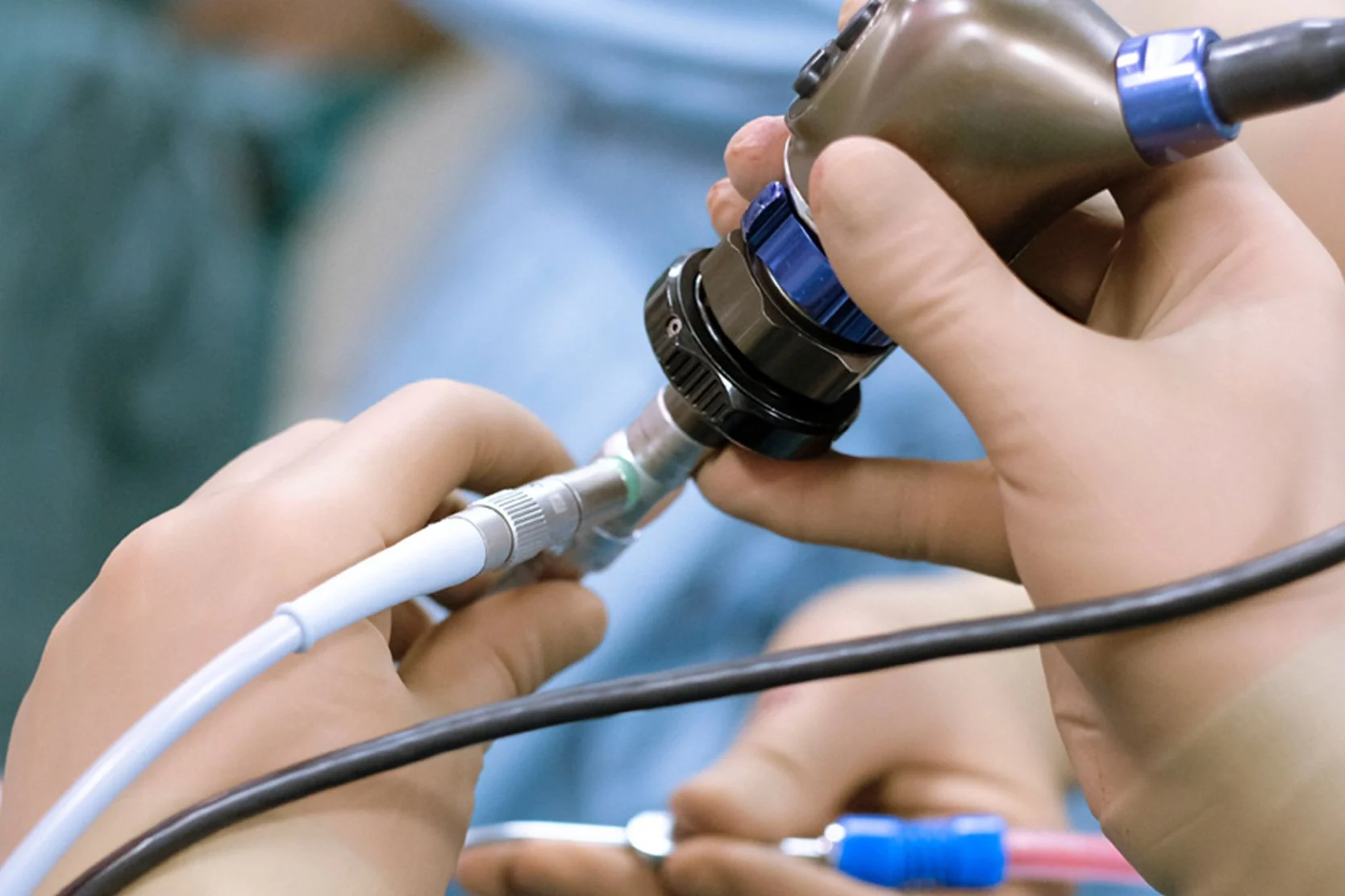
The most common treatment for gallstones is surgical removal of the gallbladder, known as cholecystectomy. UCSF gastrointestinal surgeons usually perform this as a minimally invasive laparoscopic procedure, inserting tiny instruments and a miniature camera through several small incisions.
UCSF surgeons helped pioneer laparoscopic gastrointestinal surgery, and they perform many of these procedures each year. Compared with traditional open surgery, minimally invasive surgery has significant benefits for patients, including a faster recovery, lower risk of infection, and less pain and scarring.
We also offer a nonsurgical procedure to remove gallstones from the bile duct, either during or after gallbladder removal. This option may also be used to relieve pain in patients who are too frail to undergo surgery.
Signs & symptoms
Many people do not experience any symptoms and are said to have "silent gallstones." Often the gallstones are found when a test is performed to evaluate some other problem. Treatment is only recommended if a person actually experiences symptoms of the condition.
A severe and steady pain in the upper abdomen or right side is the most common symptom of gallstones. The pain, which also may affect the shoulder blades or right shoulder, lasts anywhere from several minutes to hours. In addition, you may experience sweating or vomiting.
In its more advanced and severe stages, gallstones can cause prolonged pain and infection of the gallbladder. Stones that have passed into the bile duct usually result in pain, fever and jaundice, which is yellow discoloration of the eyes and skin.
Diagnosis
Your doctor will first ask about your medical history and perform a physical examination. In addition, he or she may order the following tests:
- Computed tomography (CT) scan. An X-ray that uses a computer to provide an image of the inside of the abdomen.
- Magnetic resonance imaging (MRI) scan. This test uses magnetic waves to create an image.
- Ultrasound. This test uses high-frequency sound waves that echo off the body to create a picture.
- Endoscopic retrograde cholangiopancreatography (ERCP). During an ERCP, a flexible tube is inserted down the throat and into the stomach and small intestine. By injecting dye into the drainage tube of the pancreas, your doctor can see the area more clearly.
- Endoscopic ultrasound (EUS). EUS involves passing a thin, flexible tube called an endoscope through the mouth or the anus to exam the lining and walls of the upper and lower gastrointestinal tract and nearby organs such as the pancreas and gall bladder. The endoscope is equipped with a small ultrasound transducer that produces sounds waves that create a viewable image of the digestive track. When combined with fine needle aspiration, EUS becomes a state-of-the-art, minimally invasive alternative to exploratory surgery to remove tissue samples from abdominal and other organs. It also may be used to determine the cause of symptoms such as abdominal pain, to evaluate a growth, to diagnose diseases of the pancreas, bile duct and gall bladder when other tests are inconclusive and to determine the extent of certain cancers of the lungs or digestive tract.
- Percutaneous transhepatic cholangiography (PTC). By injecting dye into the bile duct through a thin needle inserted into the liver, blockages can be seen on X-ray.
- Bile duct biopsy and fine needle aspiration. A tiny sample of the bile duct fluid or tissue is removed and examined under a microscope.
Treatments
Gallstones may be treated with surgery and medications.
Surgery
If surgery is required, the following procedures may be used:
- Cholecystectomy. Surgical removal of the gallbladder, a procedure called cholecystectomy, is the most widely used therapy for gallstones, although this procedure is now mostly done laparoscopically. Though in some cases, due to infections or other surgeries, this traditional form of cholecystectomy will be performed. Four or five days of hospitalization are generally required for this procedure. Patients often do well after surgery and have no difficulty with digesting food.
- Laparoscopic cholecystectomy. UCSF Medical Center also offers a less-invasive procedure called laparoscopic cholecystectomy. During this procedure, the surgeon makes several incisions in the abdomen through which a tiny video camera and surgical instruments are passed. The video picture is viewed in the operating room on a TV screen, and the gallbladder can be removed by manipulating the surgical instruments. Because the abdominal muscles are not cut there is less postoperative pain, quicker healing, and better cosmetic results. You can usually go home from the hospital within a day and resume normal activities within a few days.
- Endoscopic retrograde cholangiopancreatography (ERCP). ERCP can be used to find stones in the bile duct, as described in the diagnosis section. When stones are detected, the doctor can widen the bile duct opening and pull the stones into the intestine. This is commonly done when the gallbladder is being removed laparoscopically or when a stone is found in the duct long after gallbladder surgery. If a patient is too frail to undergo gallbladder surgery, it also may be performed to relieve symptoms from a bile duct stone, even when other stones are present in the gallbladder.
Medications
Special chemicals, available in pill form, can be used to dissolve certain gallstones, such as those composed of cholesterol. However, due to a lack of medical research, the efficacy of these medications has not been proven yet.
Awards & recognition

Best in Northern California for gastroenterology & GI surgery
More treatment info
Advanced Endoscopic Procedures
In addition to standard endoscopic procedures, UCSF Medical Center's digestive disorder specialists also offer advanced endoscopic procedures.
Support services
Case Management and Social Work
Connect with a team that can help you find resources, solve problems and advocate for you during treatment at UCSF.
Mindfulness-Based Stress Reduction Class
This eight-week class teaches mindfulness practices that can reduce stress and improve your overall health, such as meditation and body awareness.
Patient Relations
We welcome feedback about your experience at UCSF Health. Find out how to contact us with comments, questions or concerns.
Spiritual Care at UCSF
Chaplains representing many faiths are available around the clock to provide support, comfort and counsel to patients, families and caregivers.
UCSF Health medical specialists have reviewed this information. It is for educational purposes only and is not intended to replace the advice of your doctor or other health care provider. We encourage you to discuss any questions or concerns you may have with your provider.