


Brain Aneurysm

Overview
A brain aneurysm is a balloon or bubble-like growth that typically develops where a major artery branches into smaller arteries, often at the base of the brain.
Aneurysms have the potential to leak or rupture, causing bleeding into the brain or the surrounding area called the subarachnoid space. This subarachnoid hemorrhage can cause a stroke, leading to brain damage or death.
About 3 percent to 5 percent of the American population is affected by a brain aneurysm. The condition most commonly affects adults between the ages of 35 to 60 years old, although children can develop aneurysms. Aneurysms affect women more frequently than men. They can develop from continuous wear and tear on the artery walls and can be caused by factors such as genetics, injury or infection.
Our approach to brain aneurysm
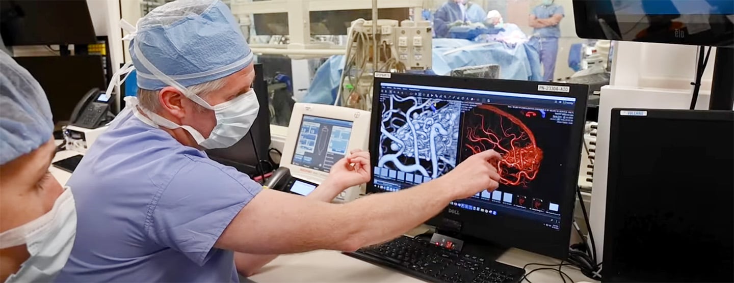
We help more than 300 patients with brain aneurysms each year – from common cases to the most complex. Our team offers advanced brain aneurysm care, including minimally invasive surgery to block the affected artery with platinum coils and bypass surgery for complex or giant aneurysms that can't be treated with conventional techniques. We were also among the first in the world to use 3D computer modeling for testing different approaches to operating on difficult-to-treat aneurysms.
Explore what we do
Options for treating brain aneurysms
Developing a brain aneurysm – a weak area in an artery that bulges with blood – can require surgery. Learn how our neurosurgical experts choose the best care.
Awards & recognition
-

Among the top hospitals in the nation
-
Best in California and No. 2 in the nation for neurology & neurosurgery
Signs & symptoms
Brain aneurysms typically don't cause symptoms until they leak or rupture, causing bleeding into the brain and are discovered during tests for other conditions. Even when they cause no symptoms, these aneurysms should be treated to prevent future ruptures.
The hallmark symptom of a ruptured aneurysm is a sudden and extremely severe headache that may occur with nausea, vomiting, stiff neck, impaired consciousness, seizures or coma.
Symptoms of an unruptured aneurysm may include:
- Cranial nerve palsy or erratic muscle movements
- Dilated pupils
- Double vision
- Localized headache
- Pain above and behind the eye
Diagnosis
A ruptured brain aneurysm is a very serious conditions that in some cases can be fatal. A quick and accurate diagnosis is essential for your recovery. Tests commonly used in the diagnosis of aneurysms include:
- Cerebral angiography. This test is commonly used to diagnose aneurysms. A special dye that can be seen on X-rays is injected into an artery that supplies blood to the brain. The dye flows through the brains blood vessels and can show any obstructions or leaks.
- Magnetic resonance angiography (MRA). Using a strong magnetic field, an MRI can generate a 3-D image of the brain and a detailed image of blood vessels that can be used to detect, diagnose and aid the treatment of aneurysms. The procedure is painless.
- Computed tomography (CT) scan. With this test, X-ray beams create a 3-D image of the brain. A CT scan can detect bleeding in the artery after the aneurysm has burst.
Treatments
Almost all brain aneurysms should be treated to prevent them from rupturing or repair ruptured aneurysms as quickly as possible and strengthen the arteries to prevent future ruptures.
Surgery
The following are some of the surgical techniques used to treat aneurysms.
- 3-D computer modeling. This technique, first performed by neurosurgeons at UCSF Medical Center, is used for difficult to treat and rare aneurysms. It produces 3-D images of the aneurysm and blood flowing through the arteries to the aneurysm. Dye is injected into arteries to track blood flow. A computer superimposes that information over brain scans to compose a 3-D model of the aneurysm. Using a computer, surgeons can test if different surgical techniques would alter blood flow enough to ease "hot spots" of pressure inside the aneurysm.
- Microsurgical clipping. A majority of aneurysms can be treated with microsurgical clipping. Aneurysms are accessed through an opening in the skull. A surgeon, using high-magnification operating microscopes, spreads apart brain tissue. A small metal clip is placed at the base of the aneurysm to tie off the bulging section of the artery. The clip must be placed without tearing the artery and causing a stroke.
- Skull base surgery. This surgery is typically used for deep and complex aneurysms located beneath the brain. The aneurysm is accessed through the bone at the base of the skull. A surgeon accesses the aneurysm from underneath or the side of the artery. The shorter pathway causes less disturbance.
- Vascular bypass grafting. Some aneurysms, such as those that are complex and large, require vascular bypass grafting. A vein is taken from the leg and used to connect an artery in the neck and an artery in the brain to bypass the aneurysm.
Endovascular therapy
A minimally invasive alternative to surgery is endovascular treatment of brain aneurysms, known as endovascular coiling. The procedure does not require an incision in the head. It is performed under general anesthesia or light sedation, and has a shorter recovery time and hospital stay compared to conventional surgery. Endovascular therapy, however, is not recommended for all patients.
The procedure involves placing small, metal coils inside the aneurysm using a catheter or long, flexible tube. The catheter is inserted into an artery in the leg and navigated through the vascular system, into the head and aneurysm.
The coils detach from the catheter and are placed in the aneurysm, where they block blood flow and cause blood to clot, destroying the aneurysm. Coils are made of platinum so they're visible on an X-ray and flexible so they can conform to the shape of the aneurysm.
UCSF Health medical specialists have reviewed this information. It is for educational purposes only and is not intended to replace the advice of your doctor or other health care provider. We encourage you to discuss any questions or concerns you may have with your provider.