


Stroke

Overview
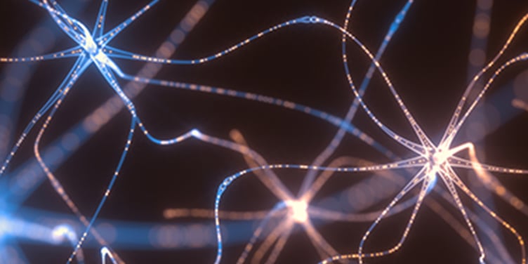
Stroke is the fifth leading cause of death and a leading cause of disability in the United States. A stroke occurs when blood flow to the brain is disrupted.
If not treated promptly, the disruption may result in permanent brain damage, long-term disability or even death. Timely treatment and follow-up care can protect brain cells and help patients recover and continue to lead healthy, productive lives.
The two main types of stroke are ischemic and hemorrhagic:
Ischemic stroke. Most strokes in the United States fall into this category. Ischemic strokes happen when a blood clot blocks the blood supply to part of the brain, preventing cells from getting oxygen and nutrients. Within a few minutes, those cells may begin to die.
The underlying cause is usually atherosclerosis, a condition in which blood vessels have become narrowed or blocked by a buildup of fatty deposits called plaque. Atherosclerosis can lead to an ischemic stroke in one of two ways:
- It can cause a cerebral thrombosis, a clot in a blood vessel leading to the brain.
- It can cause a cerebral embolism, a clot that formed in a blood vessel somewhere other than the brain (usually the heart, neck or chest) and then traveled and lodged in an artery in the brain.
Hemorrhagic stroke. This occurs when a blood vessel in the brain leaks or ruptures in one of two ways:
- Intracerebral hemorrhage. A blood vessel in the brain is bleeding into surrounding tissue. Accumulating blood can exert pressure on the brain. High blood pressure is a common cause.
- Subarachnoid hemorrhage. This is bleeding in the space between the brain and the lining that surrounds it. The cause is usually an aneurysm, a weak area in the blood vessel wall that can stretch and burst.
It's also important to be aware of a stroke-related condition called a transient ischemic attack:
Transient ischemic attack (TIA). A TIA is a stroke-like event that can cause temporary symptoms. TIAs occur when a narrowed artery or blood clot temporarily reduces the brain's blood supply. Possible symptoms include numbness, weakness or tingling in one arm or leg and problems with speech, vision or balance.
A TIA goes away in a few minutes or hours and usually doesn't cause permanent brain injury. However, it may be a warning sign of an upcoming stroke, so don't ignore the symptoms. If you have symptoms of a TIA, seek immediate medical attention. There are treatments that reduce your chance of having another TIA or a stroke.
Risk factors for stroke
Anyone can have a stroke at any age, but certain factors increase the risk of having one. Some can be treated and changed, while others are beyond your control. Even if you can't modify a risk factor, knowing about it may encourage you to take steps to reduce your overall risk.
Stroke risk factors that people can manage or modify include:
High blood pressure (hypertension). High blood pressure can damage the arteries that supply blood to the brain. Lowering it will greatly reduce your risk of having a stroke or heart attack.
Cardiovascular disease. Several heart conditions can cause blood clots and other blockages that raise stroke risk. These include coronary artery disease, a previous heart attack, congestive heart failure, a diseased aortic valve, atrial fibrillation and some congenital heart defects.
Cigarette smoking. The nicotine and carbon monoxide in cigarette smoke can damage the cardiovascular system, giving smokers almost double the risk of stroke that nonsmokers have. (Exposure to secondhand smoke also raises stroke risk.) If you smoke and take oral contraceptives, your risk of having a stroke is even higher.
Carotid artery disease. The carotid arteries supply the brain with blood. In this disorder, the vessels are narrowed by fatty deposits and can become blocked by a blood clot or produce a blood clot that leads to a stroke.
Diabetes mellitus. Having diabetes doubles the risk of stroke. Many people with diabetes also have high blood pressure, obesity and high cholesterol, all of which further raise stroke risk.
Undesirable cholesterol levels. High bloodstream levels of low-density lipoprotein (LDL) cholesterol and/or low levels of high-density lipoprotein (HDL) cholesterol can cause fatty deposits to build up in arteries, increasing stroke risk.
Obesity. Excess weight can double the risk of an ischemic stroke.
Lack of physical activity. This raises the risk of high blood pressure and therefore the risk of stroke. Being active for at least 30 minutes a day – through simple cumulative measures, such as taking the stairs instead of an elevator, or going on a daily brisk walk – can reduce your risk.
Birth control pills and hormone therapy. Women who take birth control pills, especially those who smoke and are 35 or older, have a higher risk of stroke. Women using hormone therapy for menopause are also at higher risk.
Substance abuse. Alcohol overuse can contribute to medical conditions that raise stroke risk, such as high blood pressure and diabetes. Commonly abused illicit drugs, including cocaine, amphetamines and heroin, are also associated with an increased risk, including in younger people.
Sleep apnea. A history of apnea – when breathing repeatedly stops and starts during sleep – increases the chance of having a stroke.
Risk factors that you can't control include:
Family history. The risk of having a stroke is higher if one of your parents or siblings has had a stroke.
Age. Your risk of stroke increases as you get older, doubling every 10 years after age 55.
Sex. Before age 55, men are more likely than women to have strokes. After 55, the risk is the same for men and women. However, because women tend to live longer, they're more likely than men to die of a stroke.
Previous stroke, TIA or heart attack. Once you've had a stroke, you're at greater risk for having another one. A TIA also elevates your risk: About 1 in 3 people who experience a TIA have a stroke within one year. The risk is highest during the first 48 to 72 hours after a TIA.
Race. African Americans have a higher incidence of stroke and risk of death from stroke than white Americans. Asian Americans have a higher incidence of hemorrhagic stroke than other ethnic groups.
Our approach to stroke
The UCSF Comprehensive Stroke Center offers preventive care and screening for patients at high risk of stroke, as well as the latest treatments and tools for patients who have had one. Our doctors have played key roles in developing methods for safely removing blood clots from patients experiencing a stroke. We're also one of the premier institutions for treating patients with cerebral aneurysms (weak areas in artery walls that may rupture) and subarachnoid hemorrhage.
As a world-class stroke research program, we apply the latest study findings and technologies to developing and evaluating new treatments that might improve stroke recovery and outcomes. Interested patients may have opportunities to participate in clinical trials (studies of promising treatments).
Patient stories

After a-fib and a stroke, he's back on his bike
Gary had a history of atrial fibrillation – and then a life-threatening stroke. Thanks to an embolectomy and UCSF's comprehensive care, he enjoyed a full recovery.
Stroke signs and symptoms
If you're experiencing symptoms of stroke, go to a doctor or hospital immediately, preferably by dialing 911.
Stroke symptoms generally appear suddenly (seemingly out of the blue) and can include:
- Paralysis of a leg, arm or one side of the face
- Trouble speaking or understanding speech
- Vision problems, such as blurred or double vision
- Loss of coordination or problems with balance
- Severe headache without apparent cause
- Numbness, weakness or dizziness
Temporary stroke-like symptoms can indicate a TIA. These often occur hours or days before an actual stroke, so even if the symptoms go away, you should seek medical attention right away.
Stroke diagnosis
If a stroke is suspected, your doctor will make a diagnosis based on your symptoms, medical history, physical and neurological exams, and the results of blood and imaging tests.
Your doctor will want to determine not only whether you had a stroke but the cause (a blockage or bleeding) and what part of the brain is affected. All of these factors determine your treatment.
Diagnostic tests you may undergo include:
- Blood tests. Used to check your blood sugar levels, how quickly your blood clots and whether you have an infection.
- Electrocardiogram (EKG). Measures electrical activity in the heart to detect problems that may have led to a stroke.
- Electroencephalogram (EEG). Detects abnormal electrical activity in the brain that can result from brain injury.
- Angiogram. Uses X-rays and a special dye to produce detailed images of arteries, including those in the brain. It may be performed if other tests don't reveal the stroke's cause. The doctor makes a small incision, usually in your groin, and then threads a catheter (a thin, flexible tube) through your arteries and into one of your carotid or vertebral arteries. A harmless dye is then injected into the catheter to enhance the X-ray image of your cranial arteries.
- Carotid ultrasound. High-frequency sound waves create detailed images of the interior of major neck arteries to determine whether fatty deposits are narrowing or blocking blood flow.
- Computed tomography (CT) scan. A rotating X-ray beam takes multiple images to create a detailed three-dimensional image of the brain. This shows the brain's blood vessels and blood flow, revealing areas that may be blocked or leaking blood.
- Magnetic resonance imaging (MRI). Uses a strong magnetic field to create a detailed image of the brain, allowing a specialist to detect brain tissue damaged by an ischemic stroke or a hemorrhage.
- Magnetic resonance angiography (MRA). An MRI that focuses on blood vessels. It's a noninvasive way to obtain detailed images of arteries in the neck and brain and show blockages or ruptures.
Stroke treatments
A stroke is an emergency, so head to the emergency department (ED) immediately. Certain treatments must be given within hours of the stroke to reduce (or possibly reverse) injury to the brain.
Treatment starts in the ED. You may then be admitted to the hospital for further treatment from our stroke center's inpatient service or asked to return for additional treatments as an outpatient. The kind of treatment you need depends on the type of stroke you had.
When your condition is stable, we'll address the risks that led to your stroke. For most patients, that starts with monitoring and controlling their blood pressure.
Once blood pressure is under control, we focus on getting bloodstream cholesterol to better levels, smoking cessation for patients who smoke, control of blood sugar levels for those with diabetes, and analysis of any existing heart conditions. Our neurovascular team will work with your primary care doctor to tailor treatment to your physical condition and individual needs.
Ischemic stroke
The main treatment for an ischemic stroke is a medication called tissue plasminogen activator (tPA) or a related medication called tenecteplase, which dissolves the blood clots that are blocking blood flow to the brain. It's administered through an IV (into a vein) and should be given as soon as possible – usually within four and a half hours from the time symptoms started, though some patients may be able to be treated beyond that time window.
The sooner treatment begins, the better your chances of recovery. Your doctor may also recommend one of the following procedures to treat blood vessel blockages and prevent future strokes:
- Carotid endarterectomy. If your stroke was caused by a blockage in a carotid artery, this surgery may be used to remove the plaque there. The surgeon makes an incision to open the artery, removes the plaque and closes the vessel.
- Angioplasty and stenting. Used to widen blocked vessels. The surgeon makes a small incision in the groin and threads a catheter, conveying a tiny deflated balloon, through blood vessels to the blocked artery in your neck or brain. The balloon is then inflated to widen the artery. A small, expandable tube called a stent may be permanently placed inside the widened artery to help it stay open.
- Transcarotid artery revascularization (TCAR). This minimally invasive procedure places a stent in a narrowed carotid artery to stabilize the plaque and stop it from causing a stroke.
- Thrombectomy. Used to safely remove blood clots in people having a stroke. As in angioplasty, a catheter is threaded through blood vessels. It conveys a tiny device – which UCSF doctors helped develop – that traps the blood clot and removes it.
Hemorrhagic stroke
Treatment for hemorrhagic stroke is designed to allow the brain to heal safely and prevent further bleeding. The specific treatment depends on the cause of the bleeding, its severity and which part of the brain is affected.
You may be given medications to reduce brain tissue swelling; drugs that lower blood pressure to reduce the strain on the brain's blood vessels; and vitamin K, which can stop bleeding related to certain blood thinners by helping your blood clot. Your doctor will discontinue any anticoagulant or blood-thinning medicines that may have led to bleeding.
Depending on the cause of your bleeding, you may undergo one of these procedures:
- Aneurysm clipping. To curtail bleeding from an aneurysm and prevent it from bursting again, a surgeon places a tiny clamp at its base.
- Coil embolization. To block blood flow to an aneurysm or seal it, a surgeon inserts a catheter into an artery in your groin and threads it through blood vessels to the aneurysm in your brain. A tiny coil pushed through the tube and into the aneurysm causes a blood clot to form, blocking blood flow through the aneurysm and preventing it from bursting again.
- Embolization. This technique cuts off the blood flow to an arteriovenous malformation (AVM), a tangle of arteries and veins that can break open in the brain. A surgeon fills the AVM with specially designed coils, glues or superabsorbent microspheres to plug its vessels. (Radiation may also be used to shrink an AVM.)
- Open surgery. Traditional surgery to remove an aneurysm or AVM.
- Radiosurgery. Certain types of AVMs are treated with a noninvasive tool called a Gamma Knife, which delivers a single high dose of radiation precisely to its target while causing little to no damage to surrounding tissue. The damage caused by that focused radiation seals the AVM.
If bleeding has stopped but your condition is getting worse, you may need surgery to remove clotted blood near damaged brain tissue. Or, if there's a dangerous amount of swelling, surgery may be required to reduce pressure on the brain by temporarily removing part of your skull.
UCSF Health medical specialists have reviewed this information. It is for educational purposes only and is not intended to replace the advice of your doctor or other health care provider. We encourage you to discuss any questions or concerns you may have with your provider.

Where to get care (5)
Stroke resources and support
At UCSF Health, we understand that while patients value our specialized, cutting-edge treatments, they also have emotional and practical needs related to healing. We therefore strive to provide a variety of support services to ensure that patients and families in our care are fully supported. We have a team of experienced social workers and condition-specific support groups as well as classes to help patients and families navigate their experiences while receiving care in our clinics and hospitals.
Awards & recognition
-

Among the top hospitals in the nation
-
Best in California and No. 2 in the nation for neurology & neurosurgery
-
Rated high-performing hospital for stroke
-
Certified comprehensive stroke center
Updated April 2023