Urge Incontinence in Women
On this page
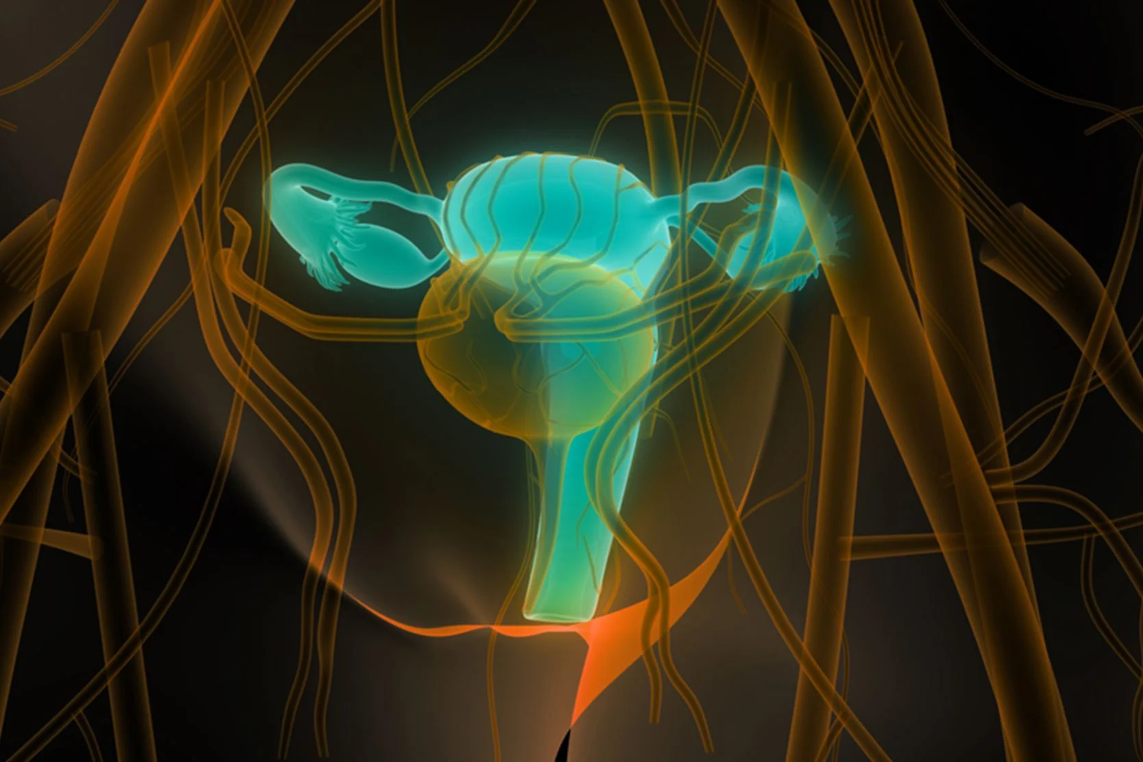
Overview
Urge incontinence is characterized by loss of urine that is associated with a sudden, strong desire to urinate that cannot be postponed. Other symptoms include a need to urinate frequently and waking often during the night to urinate. The condition is also known as overactive bladder.
Some people manage to avoid urine loss by urinating frequently, but find the continual need to have a bathroom available restrictive to their lifestyles.
Treatment for urge incontinence may include behavioral treatments such as pelvic muscle exercises, medication, electrical stimulation or Botox injections.
Our approach to urge incontinence in women
Incontinence is not an inevitable part of growing older, and there are a variety of treatments available. A leader in this field, UCSF offers innovative, compassionate care to women with urge incontinence. Our team includes gynecologists, urologists, colorectal surgeons and physical therapists who specialize in pelvic floor rehabilitation. Treatment options include bladder training, medications and Botox injections, as well as targeted exercises, biofeedback and electrical stimulation to strengthen the pelvic floor muscles. For problems with the nerves regulating the bladder, we also offer several types of nerve stimulation therapy.
We believe that empowering women with knowledge is an important part of the healing process, and encourage each patient to participate in choosing the best treatment option for her.
Signs & symptoms
The main symptom is loss of urine associated with a sudden, strong desire to urinate that cannot be postponed. Women may describe mounting pressure or sudden loss of urine in a rush to reach the toilet. Often, this occurs with certain triggering events, such as fumbling with the keys to open the front door, the sound or sensation of running water on the hands, or exposure to sudden cold.
Other symptoms include a need to urinate frequently and waking often during the night to urinate.
Diagnosis
If you have incontinence, keeping a urinary diary — a record of your daily urination, urine accidents and fluid intake — can help us make the proper diagnosis and decide on the appropriate treatment.
At your first visit to UCSF, your provider will ask questions about your general health, your history of incontinence, past surgeries, illnesses and medications you are taking. The provider will also perform a physical examination, including a pelvic exam. In addition, a urine sample will be tested. If your problem is complex, additional tests may be done at a later visit.
Physical exam assessment
Depending on the particular details of your medical history, your doctor may proceed to any or all of the following physical evaluations.
- Neurologic examination to evaluate strength, sensation and reflexes in your legs.
- Pelvic exam to assess whether you have any pelvic relaxation or pelvic organ prolapse.
- Pelvic floor assessment, in which your provider will evaluate the strength of your pelvic floor muscles, particularly your ability to contract and relax the appropriate muscle group.
- Postvoid residual urine assessment to measure how much urine remains in your bladder within 15 minutes of voiding. This test offers an estimation of your bladder's ability to efficiently "empty the tank." The same urine sample may be analyzed for other factors, such as blood, sugar, crystals or signs of infection. Such an evaluation can be accomplished with an office urine dipstick or the hospital laboratory's microscopic urinalysis.
- Urine culture if a urine dipstick or urinalysis suggests signs of acute infection. The culture is sent to the microbiology lab and, in approximately 24 to 48 hours, bacterial growth can be detected and the specific strain identified.
- Cough stress test, in which your bladder is filled with water, and you are asked to cough or strain in the same manner that would cause you to leak urine. This test can be performed in the office or incorporated into more elaborate urodynamics testing.
- A urinary diary provides details about your fluid intake and urine output, which can be crucial to making the right diagnosis. Because this is not typically the sort of information we take notice of in our daily lives, your provider will give you a urinary diary and a measuring receptacle.
You may be asked to carefully record the time and amount of any fluids you drink and the urine you void over a complete 24-hour period. You may be asked to repeat this 24-hour diary for three to five days. This allows us to notice patterns that might be important to planning your treatment.
Additional diagnostic tests
In some cases, the doctor may decide to pursue further diagnostic testing. Below is a list of some scenarios that are more complex and merit further testing:
- Uncertain diagnosis for bladder problems
- Inability to develop successful treatment plan
- Unimproved symptoms or failed treatment
- Patient is considering surgery
- Failed surgical procedure
- Prescence of other conditions, such as hematuria (blood in urine) without infection, recurrent urinary tract infections, elevated postvoid residual urine volume, neurologic condition
Urodynamic studies
The purpose of these studies is to evaluate the anatomy and function of the bladder and urethra, reproducing your symptoms.
- Cystometrogram. During this test, catheters are placed in your bladder and vagina or rectum, and your bladder is filled with fluid via the catheter. The test is used to determine your perception of water filling the bladder, any urgency to urinate, uncontrollable bladder contractions, the volume at which your bladder cannot comfortably hold any more, and the pressures within your bladder during the fluid storage process.
- Stress testing. You may be asked to perform a number of maneuvers such as coughing, changing positions, or bouncing on your heel with a catheter in place in an effort to reproduce any symptoms of incontinence.
- Urethral pressure profile. A catheter in your urethra is manipulated to measure urethral function.
- Uroflometry. During urination, a specially devised receptacle will measure the varying rate of urine flow, as well as duration of urination.
- Pressure voiding study. This test identifies abnormal voiding patterns or urine obstruction.
Other tests
In cystoscopy, a slender camera is inserted via the urethra into the bladder, enabling the doctor to view the interior anatomy of your bladder and urethra in great detail. It is typically an outpatient procedure performed for the following symptoms or situations:
- Blood or pus in urine with no bacteria present
- Bladder infections that are unusually difficult to treat
- New onset voiding irritation
- New onset bladder pain
- Suspected foreign body in the bladder
- Urodynamics tests fail to duplicate incontinence symptoms
Radiologic tests identify upper or lower urinary tract structural abnormalities:
- Intravenous pyelogram (IVP). This test involves administering intravenous (IV) dye to your bloodstream and taking X-rays of the entire urinary tract while the kidneys are processing the injected dye. This test cannot be performed if you have an allergy to IV contrast dye or abnormal kidney function.
- CT scan. A CT scan of the abdomen and pelvis may be performed, in which an X-ray machine takes a rapid sequence of two-dimensional, thin cross-sections of the body in the area of interest. This exam can be performed with or without contrast dye; mostly, this depends on what your doctor is looking for. The X-rays pictures provide great anatomic detail of most of the internal organs.
- Ultrasound. Unlike the previous two tests, an ultrasound does not involve X-rays. It utilizes a skin probe that directs sound waves to bounce off the body's internal organs, producing an anatomic picture. It can be a very useful screening tool for a number of the above indications.
Treatments
Behavioral treatments
Behavioral treatments are simple, self-directed, have no side effects and are often used in conjunction with other treatment options. They have proven effective for many women and work well for certain types of incontinence. They include:
- Bladder training. The goals of bladder training are to increase the intervals between emptying your bladder and the amount of fluid your bladder can hold. This training can help diminish the sense of urgency and leakage.
- Pelvic muscle exercises. Pelvic muscle exercises, also known as Kegels, are an essential part of improving incontinence and preventing it from worsening. They can also help you suppress the urge to urinate. The exercises strengthen and tone the muscles that support the pelvic organs. These muscles contract and relax under your command to control the opening and closing of the bladder. Achieving results requires commitment and regular exercise. Correct technique is also very important.
- Biofeedback. Biofeedback has been proven effective in numerous research studies for the treatment of urinary incontinence. It can help you learn to control and strengthen your pelvic floor muscles, which play an important role in bladder control. Because you cannot see the pelvic floor muscles, you may find it difficult to locate them or to determine if you're doing pelvic muscle exercises correctly. Biofeedback therapy uses computer graphs and audible tones to show you the muscles you are exercising. It also allows the therapist to measure your muscle strength and individualize your exercise program.
- Urge suppression. Urge suppression is a way to help control the sudden urge to urinate, so that going to the bathroom is not an emergency. Running to the bathroom is the worst thing you can do, as it increases bladder irritability and interferes with your ability to concentrate on controlling your bladder.
Medications
Medications can help relax the bladder. These medications do not cure urge incontinence, but they can be very useful in reducing or eliminating problems of bladder control. They can be used alone or in combination with behavioral treatments.
Estrogen replacement therapy taken in pill or skin patch form has not been shown to be an effective treatment for female urinary incontinence. Vaginal estrogen (creams, vaginal rings or vaginal pellets) have helped reduce recurrent urinary tract infections in postmenopausal women, but their effectiveness for incontinence is unknown.
Electrical stimulation
Pelvic floor electrical stimulation uses low-grade electrical current to stimulate weak or inactive pelvic muscles to contract. A tampon-like sensor that connects to a handheld adjustable device is inserted in the vagina. The patient then increases the current to the level of a comfortable tingle. Regular electrical stimulation sessions can supplement or augment your pelvic muscle exercise regime. Units are available for home use and may be covered by medicare or insurers.
Percutaneous tibial nerve stimulation (PTNS)
Normal voiding depends not only on the normal function of organs and muscles, but also on nerves that deliver appropriate signals regarding urination. In urge incontinence, the nerves regulating the bladder can become hyper-reactive, sending strong signals to empty before the bladder is full. Nerve stimulation therapies "jam" the pathways that transmit these abnormal messages.
In PTNS, a small acupuncture needle is placed in the ankle along the tibial nerve. A handheld device connects to the needle to deliver mild electrical impulses to the nerve. These travel up the tibial nerve to the sacral nerve plexus, which regulates the bladder. PTNS sessions are painless, last 30 minutes and are repeated weekly for 12 weeks. All sessions take place in a medical office.
Sacral neuromodulation therapy (Interstim)
Sacral neuromodulation therapy also uses electrical impulses applied directly to the sacral nerves. Wires are threaded through openings in the pelvic bones along the sacral nerves. This is done in an operating room using local anesthesia. The wires are attached to a small external generator. If a two week test using the external device shows improvement in symptoms, a permanent device, called Interstim, is implanted under the skin. This procedure requires general anesthesia.
Botox bladder injections
Botulinum toxin A, better known as botox, is used to treat patients with urge incontinence that does not improve with medications or other conservative therapies. It works by paralyzing bladder muscle, which helps decrease unwanted bladder contractions. Maximum relief is usually seen seven days after injection and normally lasts six to 12 months. Repeat injections are often needed.
Awards & recognition

One of the nation’s best for obstetrics & gynecology

Best in Northern California for urology
Recommended reading
Biofeedback for Incontinence
Biofeedback, which measures bodily processes in a way you can see, hear or understand, can help you strengthen pelvic muscles and treat incontinence.
Bladder Training
Bladder training can help treat incontinence. Get instructions to guide your bladder training so you can achieve your health goals.
Pelvic Muscle Exercises
Strengthening pelvic muscles through exercise can improve incontinence. Learn how to do pelvic muscle exercises, also known as Kegels.
Pessaries
Inserting a pessary device in the vagina can help reduce urine leakage. Learn more about pessaries, including how to insert and remove them.
Related services & treatments
Specialties
Treatments
- Pelvic Floor Exercise
- Bladder Training
- Biofeedback
- Sacral Neuromodulation for Bladder Control
Guides and forms
Support services

Case Management and Social Work
Connect with a team that can help you find resources, solve problems and advocate for you during treatment at UCSF.
Mindfulness-Based Stress Reduction Class
This eight-week class teaches mindfulness practices that can reduce stress and improve your overall health, such as meditation and body awareness.
Patient Relations
We welcome feedback about your experience at UCSF Health. Find out how to contact us with comments, questions or concerns.
Spiritual Care at UCSF
Chaplains representing many faiths are available around the clock to provide support, comfort and counsel to patients, families and caregivers.
Women's Health Resource Center
Access free health resources here, from classes and webinars to support groups and medical referrals, plus pregnancy, birth and breastfeeding services.
UCSF Health medical specialists have reviewed this information. It is for educational purposes only and is not intended to replace the advice of your doctor or other health care provider. We encourage you to discuss any questions or concerns you may have with your provider.