Kidney Cancer
On this page
Overview
What is kidney cancer?
The kidneys are a pair of bean-shaped organs located above the waist on either side of your spine. Their main job is to remove salt, waste and excess water from the blood and excrete them from the body as urine. Kidney cancer develops when some cells in a kidney begin growing out of control, eventually forming a tumor.
Our approach to kidney cancer
UCSF's urologic oncologists are internationally recognized experts in the care of people with kidney cancer. We use the latest diagnostic tools and treatments, including surgery, radiation, chemotherapy and immunotherapy (a technique that uses the body's immune system to fight cancer). Our patients have access to educational resources and support groups, as well as opportunities to participate in clinical trials evaluating new kidney cancer treatments. Learn more about the process and potential benefits of participating in a clinical trial.
Types of kidney cancer
In adults, the most common kidney cancer is renal cell cancer, or renal cell carcinoma. It accounts for 80 to 85% of kidney cancers. A less common type is transitional cell cancer. When children get kidney cancer, it's usually a form called Wilms tumor.
There are also rare types of kidney cancer and kidney tumors that are benign (noncancerous).
Renal cell cancer
Renal cell cancer occurs when cancer cells develop in the lining of small tubes inside the kidney. These tubes, called tubules, are responsible for filtering the blood and producing urine. Renal cell cancers account for 80 to 85% of all kidney cancers.
The three main types of renal cell cancer are:
- Clear cell. More than three-quarters of renal cell cancers in adults are this type. As the name suggests, the tumor cells look clear or pale when viewed under a microscope.
- Papillary. This type makes up around 15% of all renal cell cancers. The tumors are finger-like growths.
- Chromophobe. Characterized by large pale cells, this type accounts for about 5% of renal cell cancers. It tends to grow slowly compared with the other types.
Transitional cell cancer
Transitional cell cancer starts in the ureters or renal pelvis. The renal pelvis, an area at the kidney's center, collects urine and sends it to the ureters, the tubes that connect the kidneys to the bladder. This type makes up about 8% of all kidney cancers.
Symptoms of kidney cancer
In early stages, kidney cancer usually has no obvious signs or troublesome symptoms. However, as the tumor grows, it can cause blood in the urine, which may be clearly visible or present in trace amounts that can be detected in a urine test. Sometimes people can feel the tumor as a lump in the lower back or at the side of the waist.
Less common symptoms may include:
- Constipation
- Fatigue
- General feeling of poor health
- High blood pressure
- Loss of appetite
- Pain in the side or lower back that doesn't go away
- Recurrent fevers
- Unexplained weight loss
Diagnosis of kidney cancer
To begin, your doctor may ask about your medical history and perform a physical exam, carefully feeling your abdomen for any abnormal lumps. They may also recommend certain lab tests as well as imaging studies of your kidneys and nearby organs. Useful tests include:
- Urinalysis. This test involves taking a sample of your urine to assess its color and contents, measuring elements such as sugar, protein and blood cells.
- Urine cytology. This test looks specifically for cancer cells in a sample of your urine.
- Ultrasound. Using sound waves to produce images of body tissues and structures, this test is used to look for kidney tumors.
- Computed tomography (CT) scan. This special X-ray technique can produce images of the kidneys and their surrounding structures, allowing doctors to detect tumors, identify their size and location, and determine whether the cancer has spread. CT scans can also help with learning the stage of the cancer.
- Magnetic resonance imaging (MRI) scan. This imaging test uses magnets and radio waves to create high-resolution images that can show the size, location and characteristics of a tumor. It can also show the stage of cancer and whether it has spread.
- Biopsy. This test involves removing a sample of tissue from the suspicious area and examining it under a microscope for the presence of cancer cells.
Treatments of kidney cancer
Kidney cancer is usually treated with surgery, immunotherapy or both. Some patients receive radiation therapy as well. Your treatment recommendations will depend on the cancer's stage, your age and general health, and other factors. Your doctors will design a plan to fit your needs and goals.
Surgery for kidney cancer
The most common kidney cancer treatment is removing all or part of the kidney – an operation called a nephrectomy. This is an option because people can live well with just one kidney. There are two types of nephrectomies, radical and partial.
In a radical nephrectomy, the surgeon takes out the whole kidney along with the adrenal gland (a small hormone-producing gland on top of the kidney) and surrounding tissue. They may also remove some lymph nodes in the area. If the tumor is small or located on the edge of the kidney, the surgeon may remove only the part containing the tumor. This is a partial nephrectomy.
Radiation therapy for kidney cancer
Radiation therapy, also called radiotherapy, uses high-energy X-rays to kill cancer cells. Radiation therapy for kidney cancer is external, meaning that a machine outside the body delivers radioactive materials to a specific area of the body.
Patients typically receive this treatment five days a week over several weeks. The schedule is designed to protect normal tissue by spreading out the total radiation dose. You don't need to stay in the hospital for external radiation therapy, and you're not radioactive during or after treatment.
Doctors sometimes also use radiation therapy when kidney cancer has spread to the bones. In that case, it's a form of palliative therapy (comfort care) to relieve pain.
Immunotherapy for kidney cancer
Immunotherapy harnesses your body's own immune system to fight cancer. Disease-fighting cells – produced by your body or engineered in a laboratory – are used to enhance, guide or restore your body's inherent defenses against cancer. Patients typically stay in the hospital during treatment to be monitored for possible side effects.
Chemotherapy for kidney cancer
Chemotherapy is the use of drugs to kill cancer cells. Although it can treat many cancers, chemo has shown only limited effectiveness against kidney cancer. Researchers are currently studying new drugs and drug combinations with the goal of discovering some that work well for kidney cancer.
Hormone therapy for kidney cancer
Hormone therapy is used in a small number of people with advanced kidney cancer. Hormones can cause cancerous cells to divide, so this treatment may control tumor growth by targeting the cells' access to hormones. More often, it is used as palliative therapy – relieving pain or other symptoms causing discomfort.
Awards & recognition

Best in Northern California for urology

Best in California and No. 7 in the nation for cancer care
#1
in NIH funding for urology research
Helpful resources
Support services
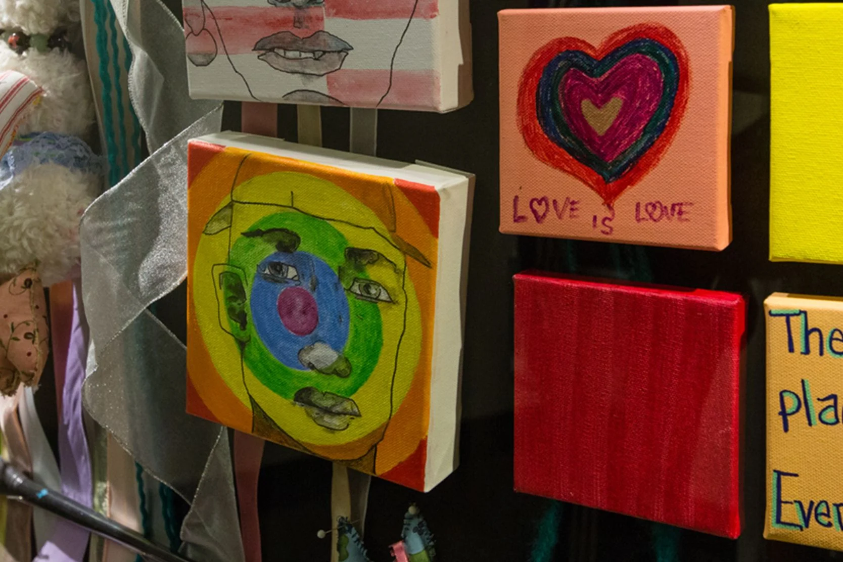
Art for Recovery
Creativity can help people with serious illnesses cope, heal and express what they're going through. Find out about our program and how to join.
Cancer Exercise Counseling
Our one-on-one exercise training sessions, customized for your needs and abilities, can complement other cancer treatments and speed your recovery.
Cancer Nutrition Counseling
UCSF Health offers free nutrition counseling to our patients with cancer, as well as nutrition seminars that are open to anyone. Learn more.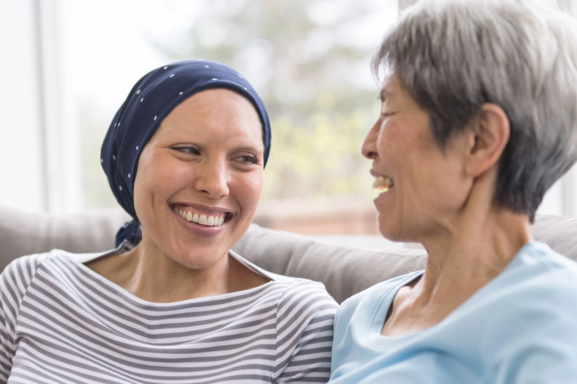
Cancer Support Groups
These groups offered by the Ida and Joseph Friend Patient and Family Cancer Support Center are free and available to all patients, whether or not you get your health care at UCSF.
Core and More Class for Cancer Patients
A strong body helps you fight cancer and enjoy life. Join this class to stabilize your core, strengthen your muscles and improve overall fitness. For cancer patients and caregivers!
Friend to Friend Specialty Shops
A one-stop boutique for patients with cancer. Get professional help with wigs, prostheses, sun-protective clothing, makeup, skin care and more.
Meditation and Guided Imagery for Cancer Patients
Drop in for a free class designed to help you heal, relax and find balance during your treatment. UCSF and non-UCSF patients are welcome.
Oncology Social Work
Social workers offer support, problem-solving, help accessing UCSF cancer-related resources and more. Find out how to contact the social worker for your clinic.
Patient & Family Cancer Support Center
The center offers wellness programming, community, support groups, classes, workshops and more at no cost to people facing cancer and their loved ones.
Peer Support Programs for Cancer
PeerTalk connects interested cancer patients with cancer survivors who can share their own experience to provide emotional and practical support.
UCSF Health medical specialists have reviewed this information. It is for educational purposes only and is not intended to replace the advice of your doctor or other health care provider. We encourage you to discuss any questions or concerns you may have with your provider.