


Aortic Stenosis

Overview
The aorta is the main artery that carries oxygen-rich blood from the heart to the rest of your body. When the valve between your heart and aorta becomes narrowed, the condition is called aortic valve stenosis.
Because the narrowed opening restricts blood flow, your heart has to work harder to pump blood through the aorta and meet your body's needs. At first, this may not cause any symptoms. As the condition progresses and the valve gets narrower, you may experience chest pain, shortness of breath, dizziness or fainting. Over time, the strain can weaken your heart and cause heart failure. Severe aortic stenosis is not preventable but can be treated.
Our approach to aortic stenosis
UCSF is internationally recognized for heart care. Our Heart Valve Disease Clinic brings together interventional cardiologists and cardiothoracic surgeons to provide comprehensive care for patients with aortic valve stenosis. Our experts offer the full range of treatments, from minimally invasive procedures to open-heart surgery. Moreover, our doctors are active in research, meaning our patients have access to the latest therapies and opportunities to take part in clinical trials (studies of promising new treatments).
Awards & recognition
-

Among the top hospitals in the nation
-

One of the nation's best in cardiology and heart & vascular surgery
Causes & symptoms
Causes
Aortic stenosis is the most common form of valve disease, occurring more often in men than women. For most people, the condition develops with age. More than 2.5 million people over the age of 75 in the United States have aortic stenosis.
Severe aortic stenosis may be related to a number of factors:
- Aging is a factor. Age-related aortic stenosis usually begins after age 60 as a result of calcium buildup in the valve, but symptoms often don't appear until a person is 70 or even 80.
- Radiation therapy, which is used to treat certain cancers, can cause thickening and calcification of the valve.
- A bacterial infection that causes scar tissue to form in the heart (as may occur with rheumatic fever) can prevent the valve from working normally.
- High cholesterol increases fat deposits, which can narrow the valve opening.
- A birth defect can affect the valve's structure and how well it functions.
Symptoms
In the mild to moderate stages of aortic stenosis, blood flow usually isn't restricted enough to cause symptoms. Many people don't know they have the condition at this stage. Some patients are told that they have a heart murmur during a routine checkup.
As the condition progresses, the opening narrows further and the heart muscle weakens. As aortic stenosis becomes severe, patients notice uncomfortable symptoms, such as shortness of breath or fatigue. This can be life-threatening, so it's crucial to tell your doctor right away if you notice new symptoms or your symptoms worsen.
Diagnosis
We use several tests to diagnose aortic stenosis and assess its severity.
Echocardiogram
An echocardiogram is an ultrasound of the heart. It produces images of the heart chambers, valves and blood flow, and can also show whether blood is clotting inside the pumping heart. We perform a transesophageal echocardiogram (TEE), in which a long flexible tube conveys a transducer (device emitting sound waves) down the throat. A TEE provides clearer images than those obtained from outside the body.
Angiogram
An angiogram is another type of imaging test. During the test, the doctor inserts a thin, flexible tube called a catheter into a blood vessel (usually in the groin area) and threads it up to the heart. The doctor then injects a harmless dye that shows up well on X-rays through the catheter to track the flow of blood to the heart.
Chest X-ray
A chest X-ray reveals the presence of calcium deposits and shows the size and shape of the heart and lungs. All patients who need heart valve surgery have a chest X-ray beforehand.
Treatment
To monitor your condition, the American College of Cardiology and American Heart Association recommend having an echocardiogram every three to five years if you have mild aortic stenosis and every one to two years if you have moderate aortic stenosis.
Your treatment depends on the severity of your condition. If the stenosis is mild or moderate, you may not have symptoms or need treatment. Be sure to tell your doctor about any new or worsening symptoms.
As the disease progresses, you may need to have the damaged valve replaced. There are several ways to do this.
Open-heart surgery
In open-heart surgery, the surgeon makes an incision in the middle of the chest to reach and remove the damaged valve and place a new one. The new valve may be made from animal tissue or a durable material, such as titanium. Or, in an open-heart operation known as the Ross procedure, one of the patient's own functional heart valves is used as the replacement. Because the heart's beating must be paused for these types of procedures, the patient is on a heart-lung bypass machine during the operation. Some patients aren't candidates for open-heart surgery because they are too ill or have other conditions that make it too risky. The Ross procedure is generally only for patients under the age of 60.
Minimally invasive valve replacement
As with open-heart surgery, the patient must be on a heart-lung bypass machine during a minimally invasive valve replacement. However, the surgeon is able to perform the procedure via a much smaller incision in the chest, using a tiny camera and slender tools to remove and replace the damaged valve. This is an option for some patients who can't have traditional open-heart surgery.
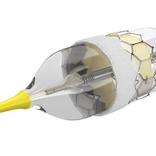
Transcatheter aortic valve replacement (TAVR)
TAVR is an even less invasive procedure we can offer to some patients, based on their evaluation. Instead of making an incision in the chest, the surgeon threads a catheter through a blood vessel to reach the heart, then inserts a new valve inside the old one. TAVR is performed while the heart is beating, so the patient doesn't need to be on a heart-lung bypass machine.
Balloon aortic valvuloplasty
This minimally invasive procedure aims to widen the narrowed valve rather than replace it. A catheter with a tiny deflated balloon at its tip is threaded through a blood vessel until it reaches the damaged valve. At that point, the balloon is inflated to expand the narrowed valve. The balloon is then deflated and the catheter is withdrawn.
UCSF Health medical specialists have reviewed this information. It is for educational purposes only and is not intended to replace the advice of your doctor or other health care provider. We encourage you to discuss any questions or concerns you may have with your provider.
More treatment info
-
Ross procedure
This surgical treatment for aortic valve disease is the only one that restores normal life expectancy to patients under 60.
Learn more -
Transcatheter aortic valve replacement (TAVR)
This minimally invasive procedure makes it possible to replace a faulty aortic valve without the risks or long recovery of open-heart surgery.
Learn more