
Soothing the soul
Our music therapy program nurtures patients with bedside serenades, rap workshops and more.
Find out more


Here to serve you
Explore our network of care for kids, from the tiniest to teens and young adults.


World-class docs
We'll help you find the best provider for your child.


Stress-free visits
Accommodations. Admission. Discharge. Procedure prep. We've got you covered.


Extraordinary kid care
Our specialists handle conditions ranging from the common to the most rare.


Referrals made easy
Contacts and resources to get your patients to our pediatric specialists


Best in Northern CA
We're ranked #1 in pediatric cancer, heart surgery, cardiology & more.


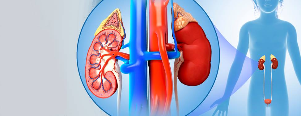
With more mothers having ultrasound scans during pregnancy, doctors are discovering more cases of hydronephrosis, a condition in which the kidney is swollen with urine due to a blocked or narrowed ureter. (Ureters are the tubes that drain urine from the kidneys into the bladder.) Before the introduction of widespread prenatal ultrasound testing, children weren't diagnosed with this condition unless they had symptoms, often after the age of 3 or 4.
Some researchers have found that up to two percent of all babies, mostly boys, have prenatal hydronephrosis. Fortunately, most of these children will never have any symptoms because the condition will either clear up or the kidneys will compensate so they work normally. But for severe or moderate cases that produce symptoms, the treatment is usually surgery.
To request an appointment, call the Fetal Treatment Center.
The three main conditions that cause hydronephrosis are:
Other conditions that can cause hydronephrosis in children include:
Children with mild and sometimes even moderate hydronephrosis usually don't have symptoms. Research suggests that the kidney compensates for hydronephrosis to maintain normal function.
However, severe hydronephrosis can damage the kidney, resulting in infections, pain and bleeding. Symptoms of urinary infection can include painful urination, cloudy urine, back pain and fever. Nephrosis, or kidney disease, can cause difficulty passing urine, either by being irregular or uncontrolled.
Hydronephrosis is usually diagnosed in one of two ways: A prenatal ultrasound reveals that the fetus has dilated kidneys, or an ultrasound that's performed to evaluate another medical problem, such as a urinary tract infection or incontinence, shows hydronephrosis. Prenatal ultrasounds detect hydronephrosis in about one out of every 100 pregnancies.
Once hydronephrosis is noted, the baby will often need additional tests to find out the severity of the condition. These tests are important because diagnosing and treating a potential abnormality early can prevent urinary tract infections and permanent kidney damage or scarring.
Tests include the following:
If your newborn child had hydronephrosis noted on a prenatal ultrasound, another ultrasound should be performed one to three days after birth. Certain conditions seen on the ultrasound, such as severe hydronephrosis in both kidneys or a dilated bladder, may warrant more tests. A VCUG will be performed within the next several weeks of life.
Hydronephrosis is graded on a scale from zero to four, with one being the mildest form and four the most severe. The degree of hydronephrosis is used to help decide how to treat the condition that's causing it. More severe grades of hydronephrosis require more extensive tests. For example, grade III and IV hydronephrosis (that are not due to vesicoureteral reflux) typically require a renal scan.
If your child's not already on antibiotics, we will give you a prescription for a low-dose, daily antibiotic. The types of antibiotics we use are very specific to the urinary tract and have very few side effects, if any. The kind of antibiotic your child receives will depend on his or her age, weight and allergies.
The antibiotics are used to prevent the hydronephrosis from causing kidney infections. Once the special X-ray tests have been completed, we can estimate how long your child will need to take the antibiotics.
The need for surgery depends on the severity of the hydronephrosis and is different for each child. Typically, non-obstructive hydronephrosis and grade I to III hydronephrosis don't need surgery and resolve over time. Children diagnosed with dilation from ureterovesical junction abnormalities called megaureters rarely, if ever, need surgical repair. Children with grade IV hydronephrosis, the most severe, are the most likely to need surgery to prevent kidney damage and recurrent infection. The surgery to correct hydronephrosis is called pyeloplasty.
UCSF Benioff Children's Hospitals medical specialists have reviewed this information. It is for educational purposes only and is not intended to replace the advice of your child's doctor or other health care provider. We encourage you to discuss any questions or concerns you may have with your child's provider.

UCSF Baskin Lab
The Baskin Laboratory investigates the causes of pediatric urological diseases present at birth or acquired later. The lab's research has a special focus on curing and preventing hypospadias, in which the urethra's opening is on the underside of the penis.

One of the nation's best in urology

Ranked among the nation's best in 10 specialties
Soothing the soul