
Definition
A mammogram is an
Alternative Names
Mammography; Breast cancer - mammography; Breast cancer - screening mammography; Breast lump - mammogram; Breast tomosynthesis; 3D mammography; 2D mammography; Diagnostic mammogram
How the Test is Performed
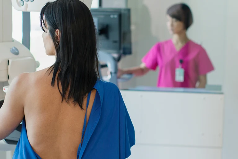
You will be asked to undress from the waist up. You will be given a gown to wear. Depending on the type of equipment used and your physical condition, you will sit or stand.
One breast at a time is rested on a flat surface that contains the x-ray plate. A device called a compressor will be pressed firmly against your breast to help flatten the breast tissue. This enables the radiologist to make a better evaluation of your breasts.
The x-ray pictures are taken from several angles. You may be asked to hold your breath as each picture is taken.
You may be asked to come back at a later date to do more mammogram images. This does not always mean you have breast cancer. Your health care provider may simply need to recheck an area that could not be clearly seen on the first test.
TYPES OF MAMMOGRAPHY
Traditional mammography uses film, similar to other x-rays.
Digital mammography is the most common technique:
- It is now used in most breast imaging centers.
- It allows the x-ray image of the breast to be viewed and manipulated on a computer screen.
- It may be more accurate in younger women with dense breasts. It has not yet been proven to help reduce a woman's risk of dying of breast cancer compared to film mammography.
Three-dimensional (3D) mammography (breast tomosynthesis) is a type of digital mammography.
How to Prepare for the Test
Do not use deodorant, perfume, powders, or ointments under your arms or on your breasts on the day of the mammogram. These substances may hide a portion of the images or produce an artifact that can falsely look like an abnormality. Remove all jewelry from your neck and chest area.
Tell your provider and the x-ray technologist if you are pregnant or breastfeeding, or if you've had a breast biopsy.
How the Test will Feel
The compressor surfaces may feel cold. When the breast is pressed down, you may feel some brief pain or discomfort. This needs to be done to get good quality images.
Why the Test is Performed
When and how often to have a screening mammogram is a choice you must make. Different expert groups do not fully agree on the best timing for this test.
Before having a mammogram, talk to your provider about the pros and cons of having the test. Ask about:
- Your
risk for breast cancer - Whether screening decreases your chance of dying from breast cancer
- Whether there is any harm from breast cancer screening, such as side effects from testing or overtreatment of cancer when it's discovered
Mammography is performed to screen women to detect early breast cancer when it is more likely to be cured. The recommendations of different expert organizations can differ.
- Mammography is generally recommended for all women starting at age 40, repeated every 1 to 2 years.
- Women with a family history of breast cancer should work with their provider to assess their risk of breast cancer. In some situations, additional testing may be considered.
Mammograms work best at finding breast cancer in women ages 40 to 74. It is not clear how well mammograms work at finding cancer in women age 75 and older.
Mammography is also used to:
- Follow a woman who has had an abnormal mammogram.
- Evaluate a woman who has symptoms of a breast disease. These symptoms may include a lump, nipple discharge,
breast pain , dimpling of the skin on the breast, changes of the nipple, or other findings.
Normal Results
Breast tissue that shows no signs of a mass or suspicious looking
What Abnormal Results Mean
Most abnormal findings on a screening mammogram turn out to be benign (not cancer) or nothing to worry about. New findings or changes must be further evaluated.
A radiology specialist (radiologist) may see the following types of findings on a mammogram:
- A well-outlined, smoothly marginated, rounded spot (this is more likely to be a noncancerous condition, such as a
cyst ) - Irregular appearing masses or lumps
- Dense areas in the breast that can be breast cancer or hide breast cancer
- Calcifications, which are caused by tiny deposits of calcium in the breast tissue (most calcifications are not a sign of cancer)
At times, the following tests are also needed to further assess mammogram findings:
- Additional mammogram views, including magnification or compression views
Breast ultrasound Breast MRI exam (less commonly done)
Comparing your current mammogram to your past mammograms helps the radiologist tell whether you had an abnormal finding in the past and whether it has changed.
When mammogram or ultrasound results look suspicious, a
Stereotactic (guided)Ultrasound (guided)Open (surgical)
Risks
The level of radiation is low and any risk from mammography is very low. If you are pregnant and need to have an abnormality checked, your belly area will be covered and protected by a lead apron.
Routine screening mammography is not done during pregnancy or while breastfeeding.
References
Allweis TM, Grubstein A, Menes T. Breast cancer screening. In: Klimberg VS, Gradishar WJ, Bland KI, Korourian S, White J, Copeland EM, eds. Bland and Copeland's The Breast: Comprehensive Management of Benign and Malignant Diseases. 6th ed. Philadelphia, PA: Elsevier; 2024:chap 12.
American Cancer Society website. American Cancer Society recommendations for the early detection of breast cancer.
American College of Obstetrics and Gynecology. Age to initiate routine breast cancer screening. ACOG Clinical Practice Update. Obstet Gynecol. 2025;145(1):e40-e44. PMID: 39388713
Henry NL, Shah PD, Haider I, Freer PE, Jagsi R, Sabel MS. Cancer of the breast. In: Niederhuber JE, Armitage JO, Kastan MB, Doroshow JH, Tepper JE, eds. Abeloff's Clinical Oncology. 6th ed. Philadelphia, PA: Elsevier; 2020:chap 88.
National Cancer Institute website. Breast cancer screening (PDQ) - health professional version.
US Preventive Services Task Force; Nicholson WK, Silverstein M, et al. Screening for breast cancer: US Preventive Services Task Force Recommendation Statement. JAMA. 2024;331(22):1918-1930. PMID: 38687503
A mammogram is a low-dose X-ray image of the breast. It can reveal small abnormalities in this tissue long before patients see or feel a change.
Mammograms are used for two main purposes: screening and diagnosis. A screening mammogram is a routine test used to detect breast cancer at its earliest stages, when it's easiest to treat. A diagnostic mammogram is a more in-depth test to investigate suspicious symptoms (such as a lump) or findings from a previous test.
Different health organizations have different guidelines for when to begin mammogram screenings and how often to get them. Many women start annual screenings at age 40, but some patients with a higher-than-average breast cancer risk may benefit from earlier or more intensive testing.
When examining your mammogram images, the radiologist will look for:
- Calcifications – These are tiny calcium deposits in breast tissue that look like white spots on the X-ray image. Certain calcification patterns can indicate breast cancer or precancerous changes. If you have an abnormal calcification pattern, your doctor may recommend a breast biopsy.
- Masses – Lumps, which doctors call masses, may be cancer but more often are caused by benign (noncancerous) breast conditions. Common benign lumps include cysts (fluid-filled sacs) and fibroadenomas (solid lumps).
Most clinics now use digital mammography rather than film. Digital mammography allows your doctor to view your images on a computer screen from multiple angles.
Digital breast tomosynthesis (DBT, also known as 3D mammography) is an advanced type of digital mammography. With DBT, we can take multiple images of a breast from different angles so the radiologist can see through overlapping tissue.
DBT is better than conventional mammography at early detection of breast cancer. DBT also produces more detailed images, resulting in fewer inconclusive mammograms that require patients to come back for additional testing.
How to prepare
To identify changes over time, your radiologist may want to compare your latest mammogram images with your past images. Learn how to send images taken at other institutions to your radiologist at UCSF Health.
On the day of your mammogram, don't use deodorant, perfume, lotion or powder on your breasts or under your arms. These products can interfere with the imaging process.
If you are pregnant or breastfeeding or have ever had a breast biopsy, make sure your doctor and mammogram technologist are aware of these things. Screening mammograms aren't performed during pregnancy or when breastfeeding.
What to expect
When you arrive, you'll be asked to undress from the waist up, change into a gown, and remove any jewelry from your neck and chest area.
Depending on the equipment being used, you either sit or stand while the images are taken. The technologist places one of your breasts on the X-ray plate. A device presses against the breast to flatten it while the image is taken. This may feel cold and uncomfortable for several seconds.
The technologist takes images from several angles and may ask you to hold your breath while each image is taken. These steps are repeated for your other breast.
The entire mammogram process usually takes about 20 minutes.
Getting the results
If your breast tissue shows no signs of masses or calcifications, your mammogram is considered normal.
If your screening mammogram reveals breast tissue changes, your doctor will want to investigate further (see "Follow-up testing," below). Most of the time, these abnormalities turn out to be benign (not cancerous). You may also be asked to come back for additional images if the radiologist couldn't clearly see any part of your breasts on the initial mammogram.
Follow-up testing
Your doctor may order the following tests to investigate suspicious findings on a mammogram:
- Additional mammogram views, including magnified images of a specific area
- Ultrasound of the breast
- MRI exam of the breast
If a mammogram or ultrasound reveals abnormalities, we may need to perform a biopsy to collect a small sample from the area of concern. A pathologist (a doctor who specializes in tissue analysis) will look for cancer cells in the sample. A biopsy is the only way to confirm a breast cancer diagnosis.
Types of biopsies include:
- Fine needle aspiration
- Ultrasound-guided core needle biopsy
- Stereotactic core biopsy
Risks
The radiation dose from a mammogram is small and doesn't significantly affect your cancer risk.
Screening mammograms aren't done when a patient is pregnant or breastfeeding. If you need a diagnostic mammogram while pregnant, the technologist will cover your abdomen with a lead apron.
Mammography myths
| Myth 1: Mammogram radiation increases my chance of getting cancer. | Fact: Modern mammography equipment uses a very small dose of radiation for each X-ray. The benefits of this procedure far outweigh the risk from this radiation dose, which doesn't meaningfully affect your cancer risk. |
| Myth 2: Mammograms don't work on younger women. | Fact: Young women tend to have denser breast tissue than older women, which can make abnormalities harder to find using standard mammography equipment. However, 3D mammography produces higher-quality images that helps doctors detect abnormalities in dense breasts. |
| Myth 3: It doesn't matter where I have my mammogram. | Fact: Studies show that radiologists who specialize in mammography, as well as those who interpret a large number of mammograms, are better at detecting cancer early. We recommend imaging centers that specialize in mammography, use the latest equipment, and are designated a breast imaging center of excellence by the American College of Radiology. If possible, use the same center each year so your radiologist can easily compare your latest mammogram to your past images. |
UCSF Health medical specialists have reviewed this information. It is for educational purposes only and is not intended to replace the advice of your doctor or other health care provider. We encourage you to discuss any questions or concerns you may have with your provider.