
Early Fetal Anatomy Evaluation
On this page
Get care
Early Anatomy Evaluation Program
We can now obtain detailed pictures of how a fetus is developing at all stages of a pregnancy, thanks to advances in prenatal imaging technologies. Our experienced team uses these tools to identify certain abnormalities as early as the first trimester.
Through our Early Anatomy Evaluation Program, we provide two types of screening, described below. Tests are conducted by our maternal-fetal medicine specialists, cardiologists, and radiologists trained in advanced prenatal diagnosis. Patients may be eligible for both types of evaluation.
Get care
Why choose UCSF
The Early Anatomy Evaluation Program is provided through the UCSF Fetal Treatment Center, which has been pioneering methods of diagnosing and treating fetal birth defects since 1981. Our patients have access to a nationally recognized team of experts in obstetrics, maternal-fetal medicine, pediatric surgery, genetics, radiology, nursing, anesthesia, social work, pediatric cardiology, pediatric cardiothoracic surgery and neonatology. For pregnancies complicated by congenital heart disease, we ensure the best care possible by working with colleagues at the UCSF Pediatric Heart Center.
As part of a children's hospital, we are able to offer comprehensive, family-centered care from diagnosis and prenatal management through postnatal care and long-term follow-up.
Early cardiac evaluation
Thanks to improved technology, heart defects and some other heart problems can be detected through a fetal echocardiogram as early as 12 to 14 weeks. Echocardiography uses sound waves to produce detailed pictures of the structure and functions of the heart as it's beating.
Early fetal echocardiography is indicated in the following circumstances:
- A suspected heart abnormality seen during the early anatomic evaluation or a routine prenatal ultrasound
- A family history of congenital heart disease
- A prior pregnancy with a fetal heart defect
- Maternal obesity or poorly controlled gestational diabetes
- Exposure to a teratogen (a substance known to cause birth defects)
- Twins or other multiple pregnancies, especially monochorionic twins (identical twins sharing a single placenta)
- A pregnancy that resulted from in vitro fertilization (IVF)
If you have any of these issues, ask your care provider for a referral. To refer a patient for early fetal cardiac evaluation, call the UCSF Pediatric Heart Center.
Early anatomic evaluation
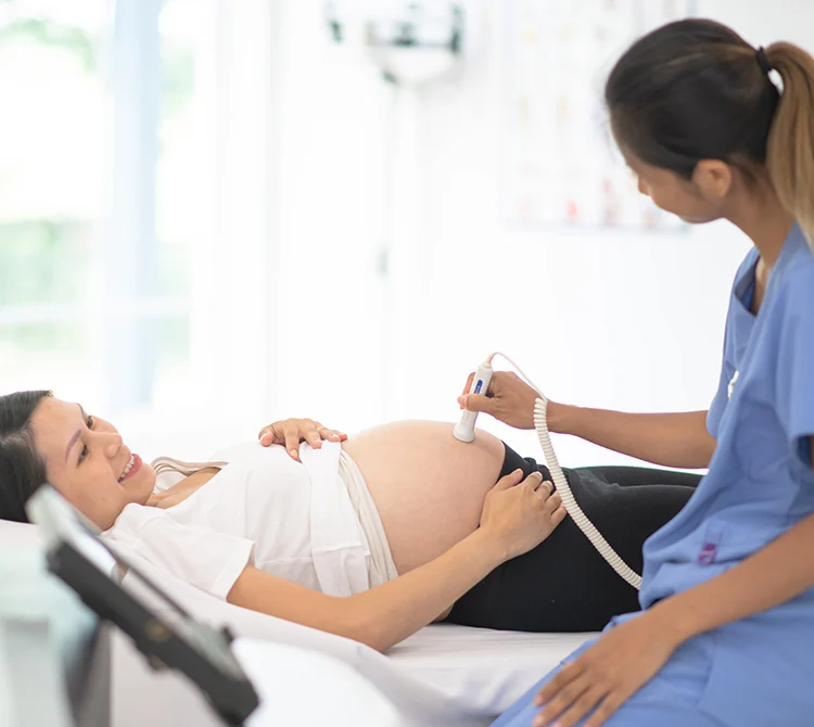
With ultrasound technology, we can perform a comprehensive evaluation of a fetus's anatomy as early as week 12 to 13 of the pregnancy. The evaluation includes brain, heart, chest, abdominal wall, stomach, kidneys and limbs.
For patients who are at higher risk for fetal abnormalities, the test provides helpful information early in pregnancy. A normal result can offer reassurance. Abnormal results give patients the chance to do follow-up imaging and diagnostic testing, so they can make informed decisions.
An early anatomic evaluation is warranted in the following circumstances:
- Suspected abnormality seen on a routine prenatal ultrasound
- Abnormal results from a cell-free DNA screening (used to check for certain chromosomal disorders)
- A family history of fetal abnormalities
- A prior pregnancy with fetal abnormalities
- Maternal obesity or poorly controlled gestational diabetes
- Exposure to a teratogen (a substance known to cause birth defects)
- Monochorionic twins (identical twins that share a single placenta)
If you're a candidate for early anatomic evaluation, ask your care provider for a referral. To refer a patient for early anatomic evaluation, call the UCSF Fetal Treatment Center.
Providers

Michael Brook, MD
Pediatric Cardiology
Katherine Connolly, MD
Maternal and Fetal Medicine
Nicole Cresalia, MD
Pediatric Cardiology • Fetal Cardiology
Neda Ghaffari, MD
Maternal and Fetal Medicine • Obstetrics and Gynecology
Related conditions & treatments
Conditions
Treatments
- Prenatal Care
- Fetal Cardiology
- Prenatal Diagnosis
- First Trimester Screening
- Prenatal Ultrasound Scan