Brain Tumor
On this page
Overview
What is a brain tumor?
A brain tumor is a mass of abnormal cells in the brain. Brain tumors can be malignant (meaning they contain cancer cells) or benign (meaning they don't contain cancer cells and won't spread to other parts of the body). There are many types of brain tumors and even benign ones can damage the brain. Early diagnosis can be an important factor in the patient's outcome. Over the next year, more than 100,000 people in the United States will be diagnosed with a brain tumor.
Our approach to brain tumors
As one of the largest and most comprehensive brain tumor programs in the U.S., the UCSF Brain Tumor Center is committed to providing our patients with the best possible outcomes and quality of life. We offer the latest treatments and techniques, such as brain mapping during surgery to protect brain function, and radiosurgery, a nonsurgical treatment that delivers high radiation doses to a precise target. Our patients also have access to clinical trials (studies evaluating promising new treatments) as well as to supportive care and resources for both them and their families.
Types of brain tumors
The two main types of brain tumors are primary and secondary.
Primary brain tumors
Tumors that begin in the brain are known as primary brain tumors and are classified by the type of tissue in which they originate. The most common brain tumors are gliomas, which begin in glial tissue (supportive tissue for nerve cells). There are several types of gliomas:
- Astrocytomas. These tumors arise from small, star-shaped cells called astrocytes. They may grow anywhere in the brain or spinal cord. In adults, astrocytomas most often arise in the cerebrum (the largest part of the brain, which is divided into right and left halves). In children, they occur in the brain stem, the cerebrum and the cerebellum (the part at the back of the head between the brain stem and cerebrum). A grade 3 astrocytoma is sometimes called anaplastic astrocytoma. A grade 4 astrocytoma is usually called glioblastoma multiforme.
- Brain stem gliomas. These tumors occur in the brain's lowest part, where it connects to the spinal cord. The brain stem controls many vital functions. Most brain stem gliomas are high-grade astrocytomas.
- Ependymomas. These tumors usually develop in the lining of ventricles, the fluid-filled cavities in the brain. They also may occur in the spinal cord. Although these tumors can develop at any age, they are most common in childhood and adolescence.
- Oligodendrogliomas. Usually located in the cerebrum, these tumors arise in the cells that produce myelin, the fatty covering that protects nerves. They are rare, grow slowly and usually don’t spread into surrounding brain tissue. They occur most often in middle-aged adults.
Some types of brain tumors do not begin in glial tissue. Among the most common are:
- Medulloblastomas. These tumors were once thought to develop from glial cells, but research has shown that they more likely develop from primitive or developing nerve cells that normally don't remain in the body after birth. For this reason, medulloblastomas are sometimes called primitive neuroectodermal tumors (PNETs). Most arise in the cerebellum, though they may occur in other areas. They most often affect children and are more common in boys than in girls.
- Meningiomas. These tumors grow from the meninges, the membranes that enclose the brain and spinal cord. They are usually benign (noncancerous). Because these tumors grow very slowly, the brain may be able to adjust to their presence and they are often quite large by the time they cause symptoms. They occur most often in women between the ages of 30 and 50.
- Schwannomas. These noncancerous tumors begin in Schwann cells, which produce the myelin that protects the acoustic nerve (the nerve responsible for hearing and balance). They occur mainly in adults. These tumors affect women twice as often as men.
- Craniopharyngiomas. These tumors develop in the region of the pituitary gland. They are usually benign but are sometimes considered malignant because they can press on or damage the hypothalamus, a small, centrally located part of the brain that regulates vital functions including body temperature, sleep-wake cycles and appetite. These tumors occur most often in children and adolescents.
- Germ cell tumors.These tumors arise from developing sex cells (germ cells). The most common type in the brain is the germinoma.
- Pineal region tumors. These tumors occur in or around the pineal gland, a tiny organ near the center of the brain. They can be slow growing (pineocytoma) or fast growing (pineoblastoma). Because the pineal region is very difficult to reach, they often cannot be removed.
Secondary brain tumors
Metastasis is the spread of cancer. When cancer begins in another part of the body and spreads to the brain, it's called a secondary tumor. Whereas primary brain tumors have their own specific types, cancer that spreads to the brain from elsewhere is the same disease and has the same name as the original (primary) cancer. For example, if lung cancer spreads to the brain, the disease is called metastatic lung cancer because the secondary tumor's cells resemble abnormal lung cells, not abnormal brain cells.
Treatment for a secondary brain tumor depends on where the cancer started and the extent of the spread, as well as such factors as the patient's age, general health and response to previous treatment.
Symptoms of brain tumors
Symptoms depend on the tumor's size and location. Symptoms are often caused by damage to vital tissue and pressure on the brain as the tumor grows within the skull's limited space.
Conditions within the brain that can lead to symptoms include fluid buildup in tissues around the tumor, a condition called edema, and hydrocephalus, which occurs when the tumor blocks the flow of cerebrospinal fluid and causes buildup in the ventricles (cavities deep inside the brain).
If a brain tumor grows very slowly, symptoms may not appear for some time. The most common symptoms of brain tumors include:
- Headaches that tend to be worse in the morning and ease during the day
- Seizures or convulsions
- Nausea or vomiting
- Weakness or loss of feeling in arms or legs
- Stumbling or lack of coordination in walking
- Abnormal eye movements or changes in vision
- Drowsiness
- Changes in personality or memory
- Changes in speech
Conditions other than brain tumors can also cause these symptoms. Diagnostic tests can help determine whether a brain tumor is the problem and, if so, whether it's a primary or secondary tumor.
Brain tumor headaches
Brain tumors can cause swelling in the brain, and the resulting pressure can lead to frequent headaches. These headaches are typically more severe in the morning and may interfere with sleep.
Diagnosis of brain tumors
To determine what's causing your symptoms, your doctor will ask about your personal and family medical history and perform a complete physical exam. In addition to assessing general signs of health, the doctor will give you a neurological exam, which includes checks for alertness, muscle strength, coordination, reflexes and response to pain. Your eyes will also be checked for swelling, which can result from a tumor pressing on the optic nerve (the nerve that sends signals from the eye to the brain).
Depending on the results of the physical and neurological examinations, your doctor may request one or both of the following:
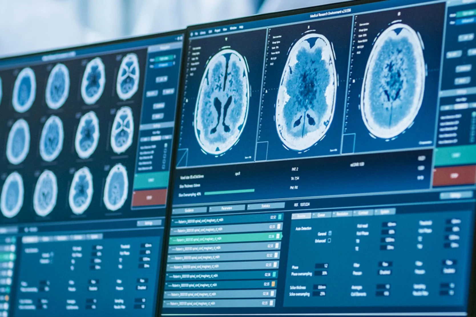
- Computerized tomography (CT) scan. This imaging method uses a computer linked to an X-ray machine to provide detailed pictures of the brain. In some cases, a special dye is injected into a vein before the scan because it can highlight abnormal tissue.
- Magnetic resonance imaging (MRI). This technology is particularly useful for brain tumor diagnosis because it provides especially detailed images of brain tissue. A contrast dye (a special dye that shows up well on imaging tests) may be used to enhance the likelihood of detecting a tumor.
The doctor may also request the following tests:
- Angiogram or arteriogram. For these tests, a contrast dye is injected into an artery. The resulting images can show the tumor and connecting blood vessels.
- Brain scan. This reveals areas of abnormal growth in the brain and records them on special film. A small amount of a radioactive material is injected into a vein. The tumor absorbs the dye and shows up on the film. The radiation leaves the body within six hours and is not dangerous.
- Functional imaging. This test utilizes magnetic resonance or magnetic source imaging to identify functional pathways in the brain (such as motor, visual and language). This information helps the surgeon avoid injuring these pathways during a procedure to remove or shrink the tumor.
- Myelogram. This test produces detailed images of your spine. A dye that shows up on X-rays or CT scans is injected into the cerebrospinal fluid. This test may be ordered when the doctor suspects a spinal cord tumor.
- MR spectroscopy. This is a modified MRI scan that shows metabolic activity within a brain tumor. Due to its superior resolution and accuracy, this technology has largely replaced positron emission tomography (PET).
Treatment of brain tumors
Brain tumor treatment options depend on several factors, including the tumor's type, location and size and the patient's age and general health. Treatment methods and schedules differ for children and adults, but in general, the methods are surgery, radiation therapy and chemotherapy. Depending on your needs, multiple methods may be used.
At UCSF, our team for brain tumor care includes neurosurgeons, medical oncologists, radiation oncologists, nurses, a dietitian and a social worker. These specialists collaborate to provide each patient with the best possible care.
Before treatment begins, most patients are given steroids, drugs that reduce swelling or edema. If you're having seizures, you may receive anticonvulsant medications to prevent or control them.
If hydrocephalus is present, you may need a shunt to drain the fluid. A shunt is a long, thin tube placed in a brain ventricle and then threaded under the skin to another part of the body, usually the abdomen. It works like a drainpipe, carrying the excess fluid from the brain to the abdomen, where it can be absorbed. In some cases, the fluid is drained into the heart.
Surgery for brain tumors
Surgery is the treatment for most brain tumors. To remove a brain tumor, a neurosurgeon makes an opening in the skull. This operation is called a craniotomy. Whenever possible, the surgeon attempts to remove the entire tumor.
If the tumor can't be completely removed without damaging vital brain tissue, your doctor may remove as much as possible. Partial removal helps relieve symptoms by reducing pressure on the brain. It also reduces the amount left to be treated through radiation therapy or chemotherapy.
Some tumors can't be removed at all. In such cases, the surgeon may do a biopsy, removing a small piece of the tumor for examination under a microscope by a pathologist. Learning the type of cells in the tumor helps your doctor decide which treatment will be most effective.
Sometimes, a biopsy is done with a needle. To pinpoint the tumor’s location, doctors use a stereotactic head frame (which facilitates precise directioning of an instrument) and CT or MRI scans. The surgeon makes a small hole in the skull and then guides a needle to the tumor.
Other advanced techniques used during surgery include:
- Brain mapping. This is to find functional pathways near the tumor so that the surgeon can avoid harming them.
- Endoscopy. A thin tube (endoscope) conveying a tiny camera can be used to perform biopsies and open pathways for draining cerebrospinal fluid.
- Frameless stereotaxic computer-assisted resection. This is an advanced method to precisely locate and remove tumors deep in the brain or in other difficult areas, such as the brain stem.
- Intraoperative MRI. Surgeons may use this imaging technique to maximize tumor removal.
Radiation therapy for brain tumors
Radiation therapy, also called radiotherapy, is the use of powerful energy rays to damage cancer cells and stop them from growing. It's often used to destroy tumor tissue that can't be removed surgically or to kill cancer cells that may remain after surgery.
Radiation therapy may be given in two ways. External radiation comes from a large machine. The treatment schedule depends on the tumor's type and size, the patient's age and other factors, but generally, external radiation treatments are given five days a week for several weeks. Giving the total dose of radiation over an extended period helps protect healthy tissue in the area of the tumor.
External radiation may be directed to the tumor only, to the tumor and surrounding tissue or to the entire brain. Sometimes the radiation also targets the spinal cord. When the whole brain is treated, the patient often receives an extra dose to the area of the tumor. This boost can come from external radiation or from an implant.
When the radiation comes from radioactive material placed directly in the tumor, this is known as implant radiation therapy, or brachytherapy. Depending on the material used, the implant may be left in the brain for a short time or permanently. Implants lose a little radioactivity each day. The patient stays in the hospital for several days while the radiation is most active.
Gamma Knife, or stereotactic radiosurgery, is another way to treat brain tumors. The Gamma Knife isn't actually a knife but a precise radiation therapy technique that delivers a finely focused, high dose of radiation to its target. Treatment is given in just one session. High-energy rays are aimed at the tumor from many angles. In this way, a lot of radiation reaches the tumor without damaging other brain tissue.
Chemotherapy for brain tumors
Chemotherapy is the use of drugs to kill cancer cells. The doctor may use just one drug or a combination, usually giving the drugs orally or by injection into a blood vessel or muscle. Intrathecal chemotherapy involves injecting these drugs into the cerebrospinal fluid.
Chemotherapy is usually given in cycles: A treatment period is followed by a recovery period, then another treatment period and so on. Patients generally don't need to stay in the hospital for treatment and most drugs can be given in the doctor's office or clinic. However, depending on the drugs used, the way they're administered and the patient's general health, a short hospital stay may be necessary.
Advances in chemotherapy include direct placement of the drug into the tumor using a new technique called convection-enhanced delivery.
Awards & recognition

Best in the West and No. 2 in the nation for neurology & neurosurgery

Best in California and No. 7 in the nation for cancer care
#1
in the U.S. for number of brain tumor patients treated
Explore what we do

Brain tumor tests lead to better outcomes
Unique program supports loved ones caring for patients with brain cancer
Recommended reading

Brain Tumor Patient and Family Resource Guide
Coping with a brain tumor can be challenging. Get resources to help you navigate the physical, emotional and financial effects of the disease.
Preparing for Gamma Knife Treatment
Here’s what to expect before, during and after Gamma Knife, an advanced radiation treatment for brain tumors, blood vessel issues and neurological conditions.
Cancer Pathology Tissue Slides FAQ
If you have a biopsy or surgery, diseased tissue is often placed on slides for a pathologist to examine. Learn how to get the slides to your doctor for review.
Cancer Radiology Scans and Reports FAQ
Learn the difference between a radiology report and radiology films or scans, why your doctor might request the scans and how to get them.
Self-Care for Caregivers
Caregiver fatigue can be brought on by the physical and emotional demands of caring for a loved one with a serious illness. Learn tips to combat caregiver fatigue here.
Communicating with Your Doctor
The doctor-patient relationship is built on communication and trust. Get tips to strengthen the partnership between you and your care team.
Coping with Chemotherapy
Everyone experiences chemotherapy differently. Understand the possible emotional and physical side effects of chemotherapy and how to cope.
Delegation to Help with Fatigue
Fatigue caused by cancer treatment can make it hard to accomplish tasks. Learn how to delegate tasks to people you trust and communicate effectively.
Nutrition Plans for Cancer Patients Undergoing Treatment
Side effects of cancer treatment may include a loss of appetite and nausea. Find out what foods to avoid and which ones may help you feel better.
Managing Your Treatment
Living with cancer or caring for someone with the disease can be overwhelming. Here are tips to reduce stress and help navigate life with cancer.
Nutrition and Coping with Cancer Symptoms
Side effects of cancer treatment may affect your eating habits. Find new ways to get the calories, protein and nutrients you need.
Questions to Ask Your Cancer Doctor
These questions can guide conversations with your cancer doctor and help you get the information you need to make care decisions for yourself or a loved one.
Resources for End of Life
Get resources to support yourself and your loved ones through end-of-life issues. Find bereavement support groups, counselors and reading materials.
Tips for Conserving Your Energy
Cancer treatments can cause extreme exhaustion. Here are tips to help you conserve your energy and manage cancer-related fatigue.
Using a Medical Calendar and Symptom Log
Here’s how to keep a medical calendar and symptom log to help you and your doctor track your condition and side effects of treatments or medications.
More treatment info
Brain Mapping
The brain is the most complicated organ in our body. Every area has a specific function that controls everything that we do. Find more information here.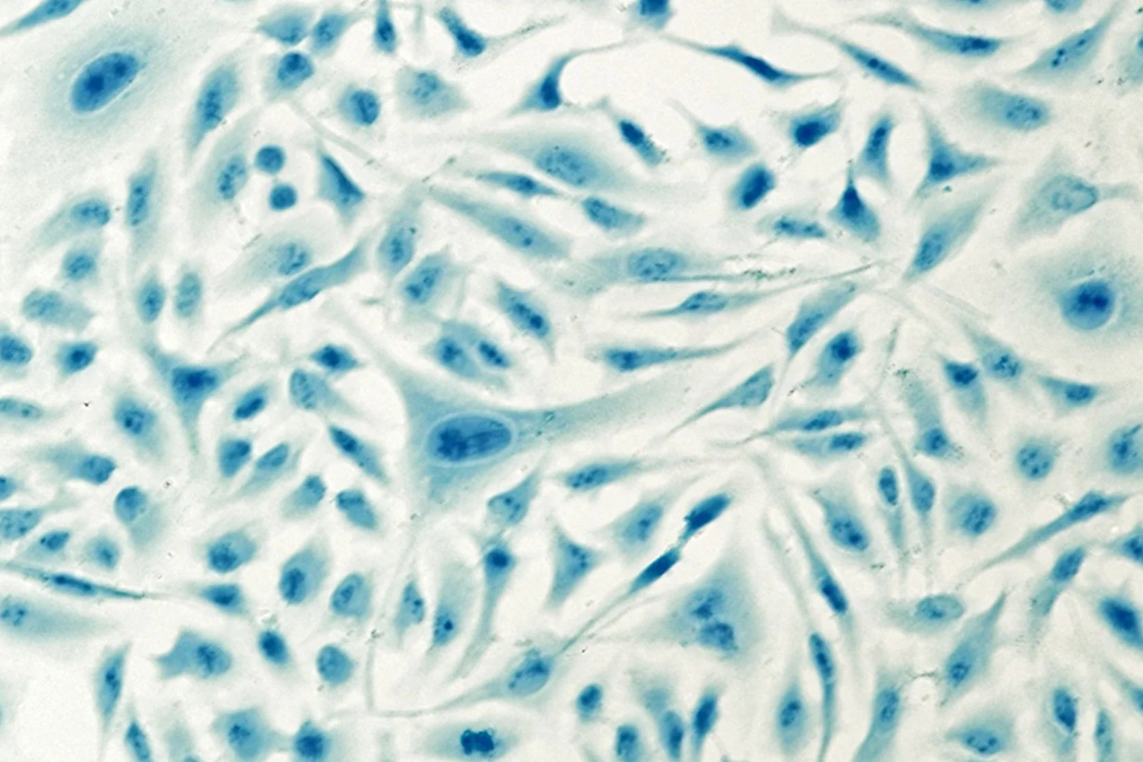
Brachytherapy
Brachytherapy delivers radiation by placing radioactive material either directly inside a tumor or very close to it, sparing healthy tissue.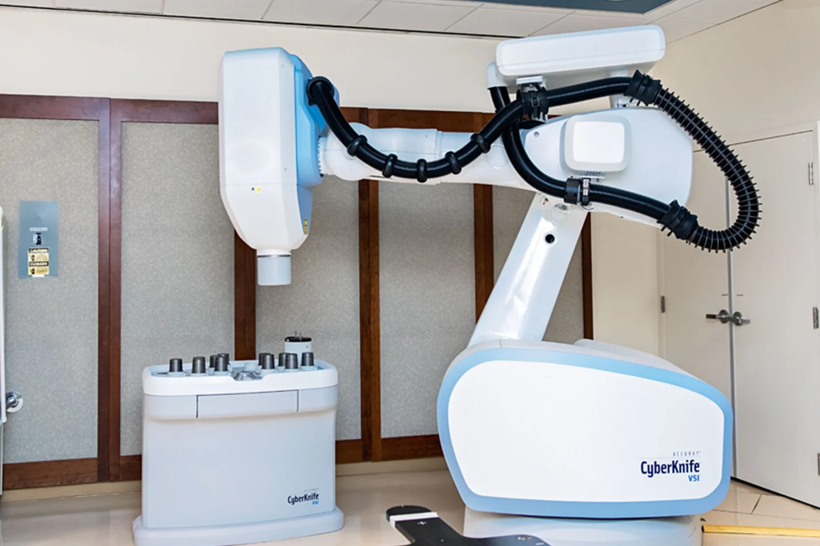
Cyberknife
The CyberKnife one of the most advanced forms of radiosurgery is a painless, non-invasive treatment that delivers high doses of precisely targeted radiation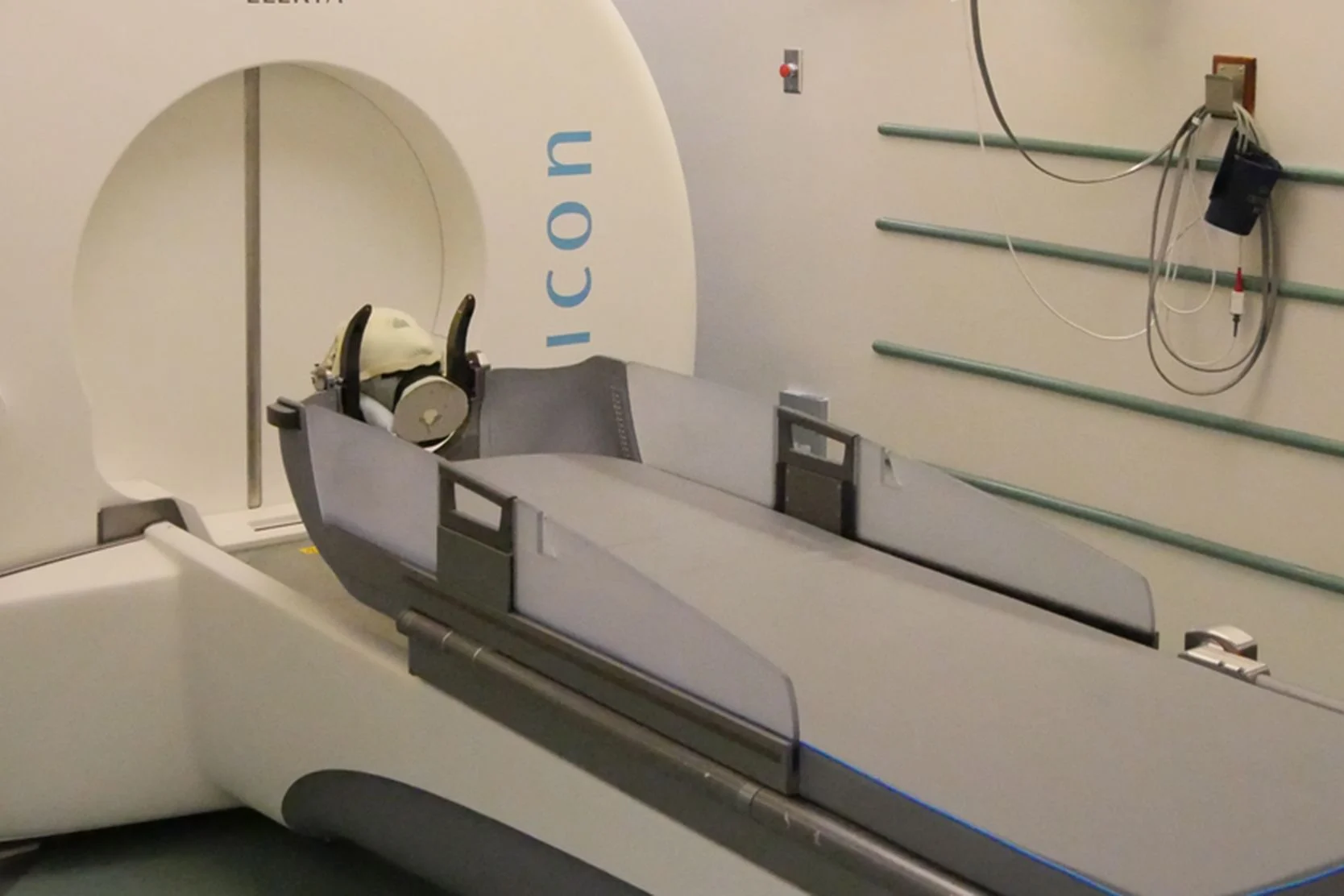
Gamma Knife
The Gamma Knife is an advanced radiation treatment for adults and children with small to medium brain tumors, abnormal blood vessel formations, and more.
Support services

Art for Recovery
Creativity can help people with serious illnesses cope, heal and express what they're going through. Find out about our program and how to join.
Cancer Exercise Counseling
Our one-on-one exercise training sessions, customized for your needs and abilities, can complement other cancer treatments and speed your recovery.
Cancer Nutrition Counseling
UCSF Health offers free nutrition counseling to our patients with cancer, as well as nutrition seminars that are open to anyone. Learn more.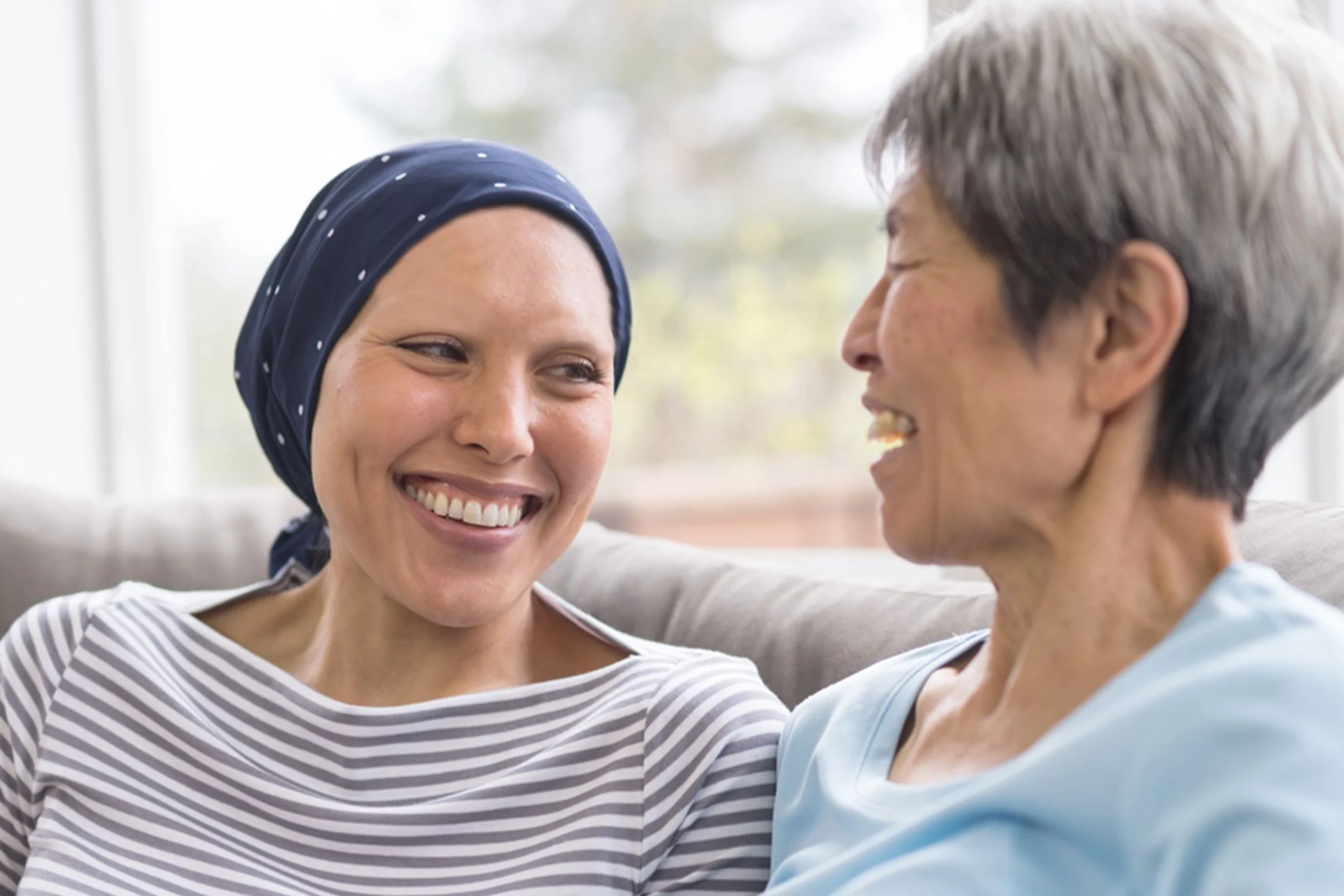
Cancer Support Groups
These groups offered by the Ida and Joseph Friend Patient and Family Cancer Support Center are free and available to all patients, whether or not you get your health care at UCSF.
Core and More Class for Cancer Patients
A strong body helps you fight cancer and enjoy life. Join this class to stabilize your core, strengthen your muscles and improve overall fitness. For cancer patients and caregivers!
Friend to Friend Specialty Shops
A one-stop boutique for patients with cancer. Get professional help with wigs, prostheses, sun-protective clothing, makeup, skin care and more.
Meditation and Guided Imagery for Cancer Patients
Drop in for a free class designed to help you heal, relax and find balance during your treatment. UCSF and non-UCSF patients are welcome.
Neuro-Oncology Caregiver Program
If you're caring for someone with a brain tumor, our program can help with guidance, information, resources and support at this difficult time. Find out more.
Oncology Social Work
Social workers offer support, problem-solving, help accessing UCSF cancer-related resources and more. Find out how to contact the social worker for your clinic.
Patient & Family Cancer Support Center
The center offers wellness programming, community, support groups, classes, workshops and more at no cost to people facing cancer and their loved ones.
Peer Support Programs for Cancer
PeerTalk connects interested cancer patients with cancer survivors who can share their own experience to provide emotional and practical support.
UCSF Health medical specialists have reviewed this information. It is for educational purposes only and is not intended to replace the advice of your doctor or other health care provider. We encourage you to discuss any questions or concerns you may have with your provider.