Breast Cancer
On this page
Overview
What is breast cancer?
Breast cancer is the second most common cancer among women, with more than 200,000 new cases diagnosed each year nationwide. While known primarily as a women’s disease, men can also get breast cancer.
Breast cancer begins with abnormal cells developing in breast tissue. The cancer may be confined to the breast or spread to other parts of the body. The most common type begins in the ducts that carry milk to the nipple. But cancer also may occur in the small sacs that produce milk, called lobules, or in other breast tissue. Because breast cancer varies widely, treatment depends on the type and stage as well as the individual patient's needs.
Our approach to breast cancer
Breast cancer survival rates today are higher than ever due to advances in diagnosis and treatment. At the UCSF Carol Franc Buck Breast Care Center, we specialize in screening to enable early detection, providing more effective and less toxic therapies, educating patients and pursuing cutting-edge research. We are committed to discovering new and better ways to prevent and treat breast cancer.
UCSF offers the highest quality breast cancer care in a respectful and supportive environment. To serve each of our patients, our surgeons, oncologists, radiologists, psychologists and nutritionists work as a team to heal the whole person, both physically and emotionally.
Types of breast cancer
Breast cancer may be found in several forms, depending on where the cancer started and how far it has progressed.
Breast cancer in situ, DCIS and LCIS
When breast cancer is found early, usually through a mammogram, it's called breast cancer in situ (meaning in its original position) or noninvasive cancer. However, it can develop into invasive breast cancer.
Two types of breast disease in situ are:
- DCIS (ductal carcinoma in situ). Abnormal cells are only in the lining of a milk duct (they haven't spread beyond the duct). If these cells aren't removed, some may change over time and become invasive cancers. DCIS is sometimes called intraductal carcinoma.
- LCIS (lobular carcinoma in situ). Abnormal cells are found only in the lining of a milk lobule. LCIS is noninvasive and not considered breast cancer. However, it signals an increased risk of developing invasive cancer. LCIS is sometimes found in a biopsy for another lump or when an unusual change is detected on a mammogram.
Invasive breast cancer
When cancer cells form in the ducts or milk lobules and spread to the breast tissue around them, the cancer is considered invasive. Tumors can be found during a breast exam or through screening tests, such as a mammogram. The severity of the cancer and best course of treatment depend on the tumor's size, what the cells look like under a microscope, and whether the cancer has spread to lymph nodes (small, bean-shaped structures that are part of the body's immune system).
Metastatic breast cancer
The cancer is metastatic when it has spread from the breast to other organ systems – usually the bones, lungs, liver and brain. The cancer travels to these organs through the bloodstream or lymphatic system. If metastatic cancer develops, it is usually months or years after the initial breast cancer diagnosis.
Inflammatory breast cancer
Inflammatory breast cancer is a rare but aggressive type. The breast may look red and feel warm. Ridges, welts or hives may appear on the breast, or its skin may look wrinkled. Inflammatory breast cancer is sometimes misdiagnosed as an infection.
Recurrent breast cancer
This is cancer that has come back (recurred) after treatment. It may reappear in the breast, develop in soft tissues of the chest or chest wall, or occur in another part of the body.
Symptoms of breast cancer
Early in the development of breast cancer, people don't usually experience pain. In fact, when breast cancer first appears, there may be no symptoms. But as the cancer grows, it may cause changes. Watch for differences in appearance or sensation, including:
- A lump or thickening in or near a breast or the underarm area
- A change in the breast's size or shape
- Nipple discharge or tenderness
- The nipple pulling inward (inverting)
- Ridges or pitting of a breast, making the skin look like the outside of an orange
- Any change in the look or feel of the skin of a breast, areola or nipple, such as warmth, swelling, redness or scaliness
Diagnosis of breast cancer
To diagnose breast cancer, your provider will usually perform a physical exam and order an imaging test, such as a mammogram or ultrasound. Depending on the results, you may also have a tissue biopsy (a procedure to take a sample for microscopic analysis).
If cancer is found in your breast, you'll undergo additional tests to determine the stage of the disease. Staging is a way of assessing whether the cancer has spread and, if so, to which parts of the body. Blood and imaging tests are typically used. Your treatment plan will depend on the results of these tests as well as your own needs and preferences. You can find more information on the staging of breast cancer in Basic Facts About Breast Health.
Imaging for breast cancer
Imaging tests are used to diagnose breast cancer and to evaluate its stage and extent. Based on the results, your provider may recommend further tests or therapy – or determine that no treatment is necessary.
Imaging tests may include:
- Screening mammogram. A mammogram is a low-dose X-ray of the breast. This is the best test to check for breast cancer in people without signs of it.
- Diagnostic mammogram. This type of mammogram is used to investigate suspicious changes in the breast, such as a new lump or breast pain or nipple discharge. It's also used to evaluate suspicious findings on a screening mammogram.
- Breast ultrasound. This test uses high-frequency sound waves to show whether a lump is solid or filled with fluid. It may be used along with diagnostic mammography or an MRI to answer questions about a specific area of the breast.
- Breast magnetic resonance imaging (MRI). This type of MRI scan can reveal abnormalities that aren't visible through mammography or ultrasound. Each scan produces hundreds of images.
The American Cancer Society recommends that certain women with an especially high risk of developing breast cancer have an MRI scan along with their yearly mammogram. A breast MRI is noninvasive, with no radiation exposure. But breast MRI is an evolving technology and shouldn't replace standard screening and diagnostic procedures, such as in-clinic exams during a regular checkup, self-exams, mammograms and biopsies.
Biopsy for breast cancer
A biopsy may be used to determine whether a breast lump or other abnormal tissue is cancer. During a biopsy, a surgeon, pathologist or radiologist removes a portion or all of the suspicious tissue. The tissue is then examined under a microscope by a pathologist, who checks for cancer cells and makes the diagnosis.
Types of biopsies include:
- Fine-needle aspiration (FNA) biopsy. Using a small, thin needle, the doctor takes two or three samples of tissue from the breast lump for examination.
- Stereotactic core biopsy. Using a biopsy needle, the doctor removes tissue from your breast while it’s pressed down to hold it in position, similar to what happens during a mammogram.
- Magseed or needle (wire) localization biopsy. Guided by imaging, doctors mark the location of the abnormal tissue by placing a tiny magnetic seed or wire. The tissue is then surgically removed and analyzed.
Decision-making consultation
If you're diagnosed with breast cancer, the Patient Support Corps at the UCSF Breast Care Center is here to help. The staff can offer information and guidance as you navigate conversations with your doctors and make decisions about your treatment.
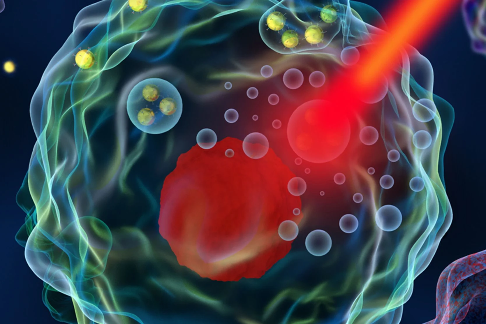
Treating breast cancer
Treatment for breast cancer may include surgery as well as radiation, chemotherapy and hormone therapy. Local treatments, such as surgery and radiation therapy, remove, destroy or control cancer cells in specific areas. Systemic treatments, such as chemotherapy and hormone therapy, destroy or reduce cancer throughout the body.
Depending on your condition, you may receive one treatment, a combination of therapies at the same time or a series over time.
Surgery for breast cancer
For most types of breast cancer, surgery is the most common treatment. Mastectomy and lumpectomy are two main surgeries for removing tumors. Additional procedures may be done to check for cancer in your lymph nodes or to reconstruct your breast during or after removal.
Your doctor may discuss these procedures with you:
- Lumpectomy. A surgeon removes the cancer and some normal tissue around it, preserving as much healthy tissue as possible. Some lymph nodes from the armpit may also be removed to determine whether the cancer has spread. After a lumpectomy, most patients receive radiation therapy to destroy any remaining cancer cells.
- Mastectomy. A surgeon removes all the tissue of one or both breasts. Other nearby tissues, such as lymph nodes, may be removed at the same time to check whether the cancer has spread.
- Sentinel lymph node biopsy. A surgeon removes one or more sentinel lymph nodes for examination. Sentinel lymph nodes are the ones most likely to contain cancerous cells if the cancer has spread from the primary area.
- Breast reconstruction. A surgeon rebuilds the breast after a mastectomy, creating a breast that looks and feels as natural as possible. This procedure may be done using breast implants or the patient's own body tissue, although a variety of factors affect an individual's options.
Radiation therapy for breast cancer
Radiation therapy for breast cancer uses high-energy rays or particles to attack the disease. This treatment is delivered in effective, tolerable doses to kill tumor cells or inhibit their growth and division.
Radiation therapy is used, along with surgery, to treat early-stage breast cancer. It may also be used in more advanced breast cancer to control the disease or to relieve symptoms, such as pain.
Chemotherapy for breast cancer
Chemotherapy uses drugs to kill cancer cells. For breast cancer, the drugs are typically a combination given by mouth and injection. Chemotherapy enters the bloodstream and travels throughout your body.
Chemotherapy is most commonly used to:
- Decrease the chance of cancer recurring after surgery
- Shrink breast cancer after surgery when the tumor is large or inflammatory
- Control metastatic breast cancer that has spread to other organs
Hormone therapy for breast cancer
This therapy changes the hormonal environment in your body, affecting the growth and behavior of some breast cancers. If your breast cancer produces an estrogen receptor or progesterone receptor, hormone therapy can effectively treat it at early, metastatic or advanced stages. This therapy can also be used to prevent recurrence.
Awards & recognition

Best in California and No. 7 in the nation for cancer care
Designated comprehensive cancer center
Recommended reading

Basic Facts About Breast Health
Learn about breast health, including breast anatomy, breast cancer screening and lifestyle changes to help prevent breast cancer.
Biopsy for Breast Cancer Diagnosis: Fine Needle Aspiration
Fine needle aspiration biopsy (FNA) is a quick procedure your provider may use to identity if a breast lump is cancerous. Here’s what to expect from an FNA.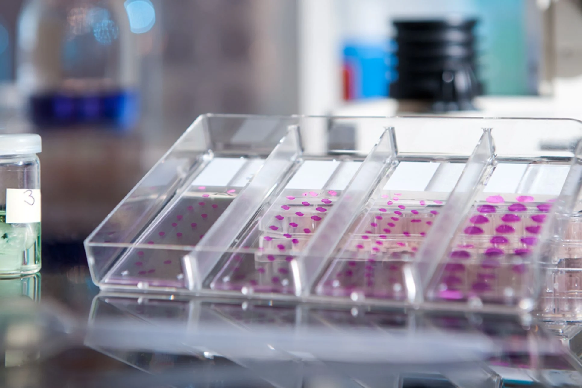
Biopsy for Breast Cancer Diagnosis: Magseed and Needle (Wire) Localization
Learn more about magseed and needle (wire) localization to diagnose breast cancer, including how to prepare, procedure steps, and risks and recovery.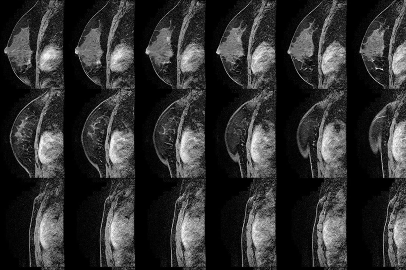
Biopsy for Breast Cancer Diagnosis: MRI-Guided Core Biopsy
During an MRI-guided core biopsy, your team collects a small sample of breast tissue to examine. Here’s what to expect before, during and after the procedure.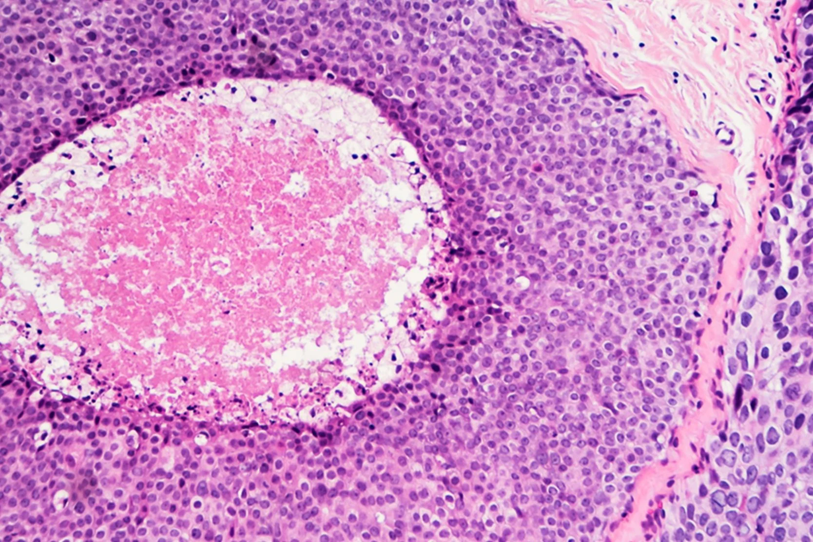
Biopsy for Breast Cancer Diagnosis: Surgical Breast Biopsy
Understand the surgical biopsy process for breast cancer diagnosis. Learn how to prepare for breast biopsy surgery and what to expect during recovery.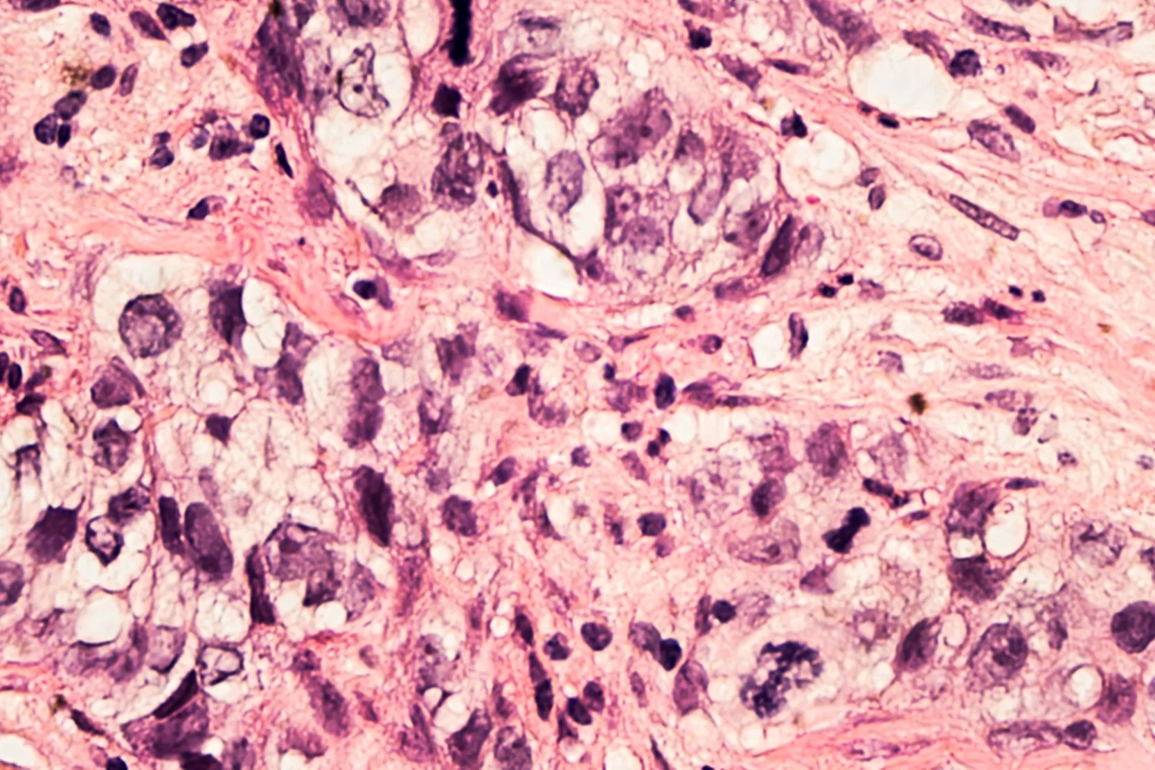
Biopsy for Breast Cancer Diagnosis: Stereotactic Core Biopsy
We may use stereotactic core biopsy to collect a small sample of breast tissue. Find out what happens during the procedure and when you can expect results.
Breast Cancer Glossary
This comprehensive glossary of breast cancer terminology includes helpful definitions of everything from AC chemotherapy to X-ray.
Breast Cancer Risk Factors
Some people have a higher risk of developing breast cancer than others. Find out what factors increase risk, including those we can and cannot change.
Follow-Up Care for Breast Cancer Patients
After completing early-stage breast cancer treatment, here’s what to expect for follow-up exams, mammograms and other screening tests.
Preparing for a Lumpectomy or Mastectomy
Here’s how to prepare for a lumpectomy or mastectomy, including arrangements you should make before surgery, what to eat and items to bring to the hospital.
Menopause and Breast Cancer
Breast cancer treatment often causes premature menopause and can trigger certain side effects. Find out how to deal with menopause symptoms.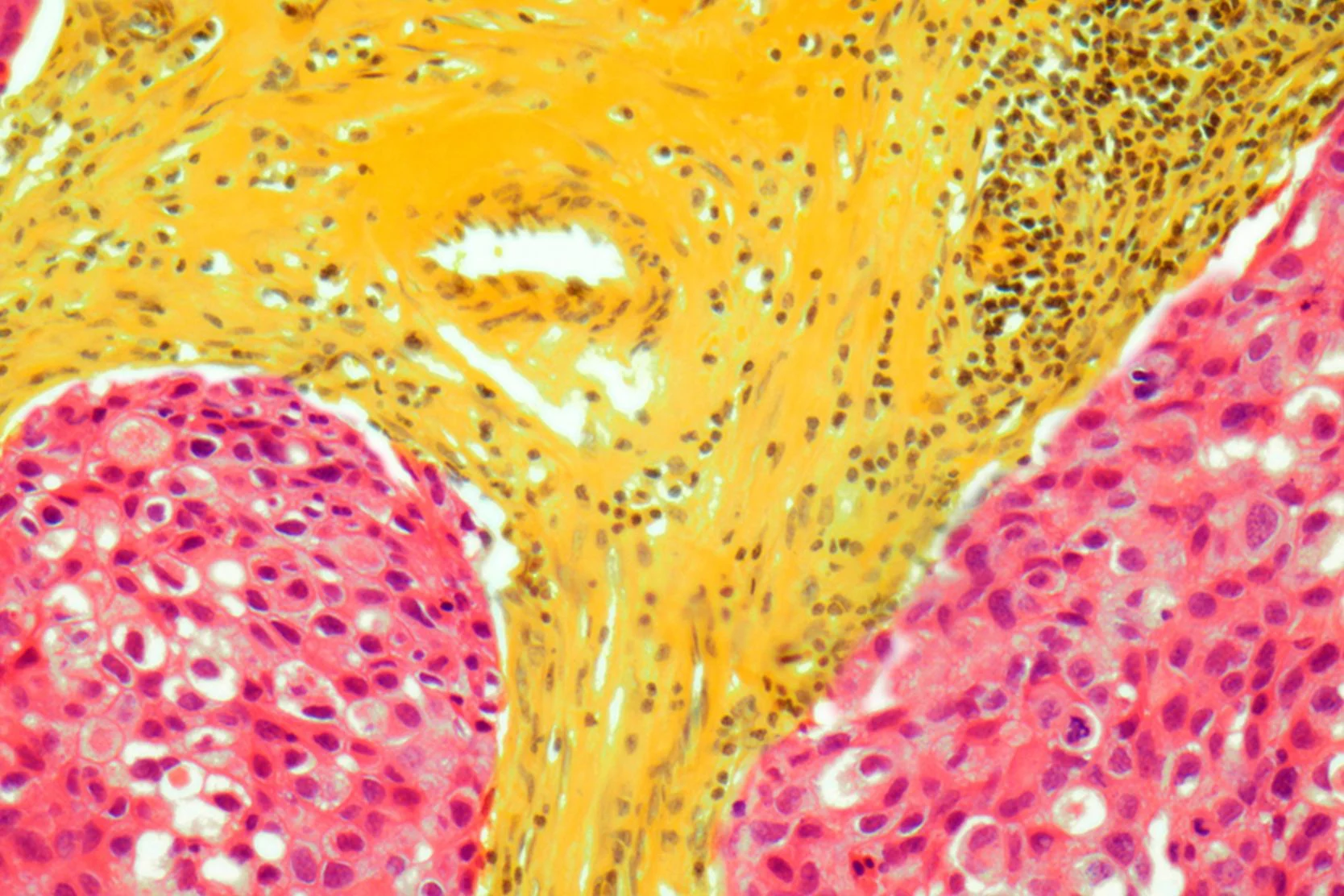
Metastatic Breast Cancer: Diagnosis and Treatment
Metastatic breast cancer is cancer that originated in the breast and has spread to other parts of the body. Learn what may affect treatment options.
Navigating Breast Care
No matter what kind of breast care you need, you can find resources and guidance to support your breast health.
Osteoporosis and Breast Cancer
Having had breast cancer increases your risk of developing osteoporosis. Find out how we diagnose osteoporosis using a bone mineral density (BMD) test.
Radiation Therapy for Breast Cancer
Learn about radiation therapy for breast cancer, including what to expect during treatment and how to care for yourself between sessions.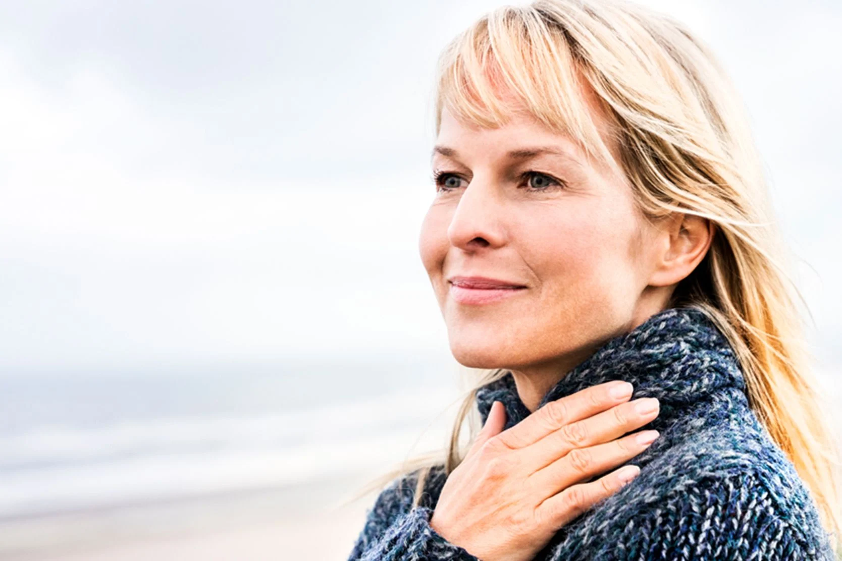
Self-Care and Recovery
Self-Care and recovery resources including an Introduction to Lifestyle Change, Nutrition and Breast Cancer, Hydration: Water and Health, Meditation and more.
Taking Charge: Women's Health
Knowledge about female cancers can help you take action and protect yourself. Get the facts about breast and ovarian cancers.
Related programs

UCSF Patient Support Corps
Receiving a breast cancer diagnosis is stressful. Get guidance, support and answers to your questions here as you consider your options and make decisions.
More treatment info
Hyperthermia
Hyperthermia is a treatment, performed by Radiation Oncology, that uses very high heat levels to kill small cancer tumors and lower levels to enhance radiation.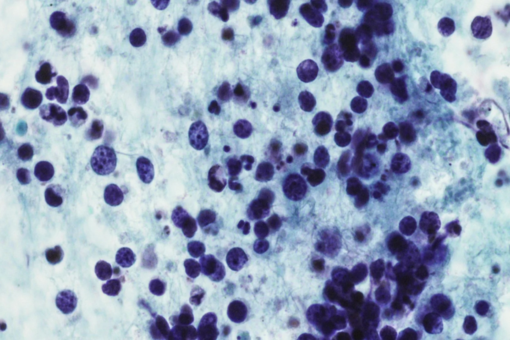
Intensity Modulated Radiation Therapy
IMRT is the most advanced form of 3-D conformal RT, allowing for higher, more effective doses of radiation.
Lumpectomy
A lumpectomy, or partial mastectomy, is a surgical procedure for breast cancer designed to preserve as much normal tissue as possible.
Mastectomy
Mastectomy is the surgical removal of breast tissue. Learn about various mastectomy techniques and find resources to help you prepare.
Helpful resources
Support services

Art for Recovery
Creativity can help people with serious illnesses cope, heal and express what they're going through. Find out about our program and how to join.
Cancer Exercise Counseling
Our one-on-one exercise training sessions, customized for your needs and abilities, can complement other cancer treatments and speed your recovery.
Cancer Nutrition Counseling
UCSF Health offers free nutrition counseling to our patients with cancer, as well as nutrition seminars that are open to anyone. Learn more.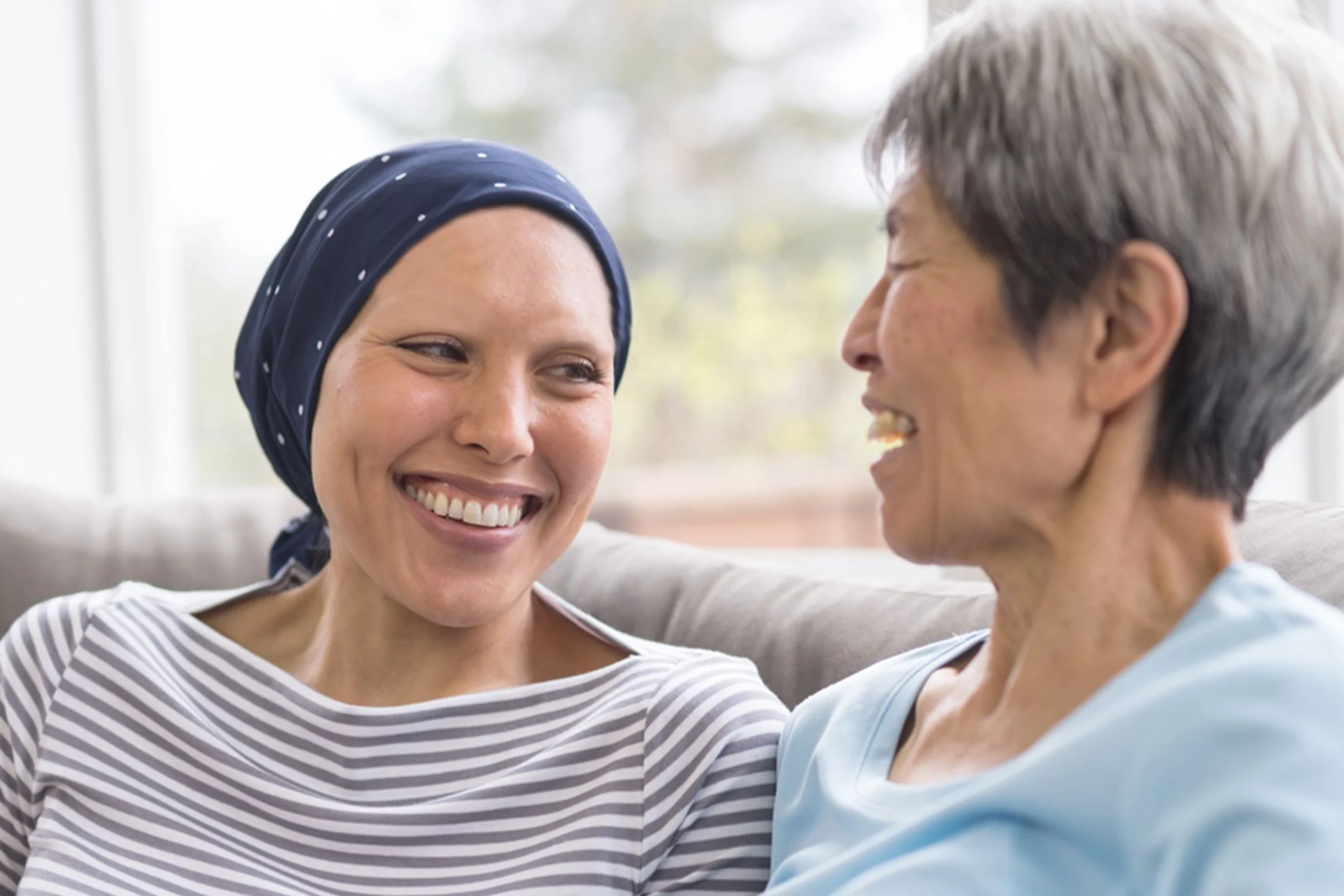
Cancer Support Groups
These groups offered by the Ida and Joseph Friend Patient and Family Cancer Support Center are free and available to all patients, whether or not you get your health care at UCSF.
Core and More Class for Cancer Patients
A strong body helps you fight cancer and enjoy life. Join this class to stabilize your core, strengthen your muscles and improve overall fitness. For cancer patients and caregivers!
Friend to Friend Specialty Shops
A one-stop boutique for patients with cancer. Get professional help with wigs, prostheses, sun-protective clothing, makeup, skin care and more.
Meditation and Guided Imagery for Cancer Patients
Drop in for a free class designed to help you heal, relax and find balance during your treatment. UCSF and non-UCSF patients are welcome.
Oncology Social Work
Social workers offer support, problem-solving, help accessing UCSF cancer-related resources and more. Find out how to contact the social worker for your clinic.
Patient & Family Cancer Support Center
The center offers wellness programming, community, support groups, classes, workshops and more at no cost to people facing cancer and their loved ones.
Peer Support Programs for Cancer
PeerTalk connects interested cancer patients with cancer survivors who can share their own experience to provide emotional and practical support.UCSF Patient Support Corps
Receiving a breast cancer diagnosis is stressful. Get guidance, support and answers to your questions here as you consider your options and make decisions.
UCSF Health medical specialists have reviewed this information. It is for educational purposes only and is not intended to replace the advice of your doctor or other health care provider. We encourage you to discuss any questions or concerns you may have with your provider.